
Overview
Age-related macular degeneration (AMD) is a progressive disease characterised in its earlier stages by the presence of drusen and/or pigmentary changes. Drusen are extracellular deposits that lie between the RPE and Bruch's membrane. They can vary significantly in size and distribution and can be seen in various conditions outside of AMD as well.
In AMD, drusen typically increase in size and number over time. However, drusen regression can also occur which may signal an increased risk of progression to late AMD.
Several different types of drusen can be identified using multimodal imaging. The imaging characteristics of each are illustrated below.
Drusen Variants
-
Drusen
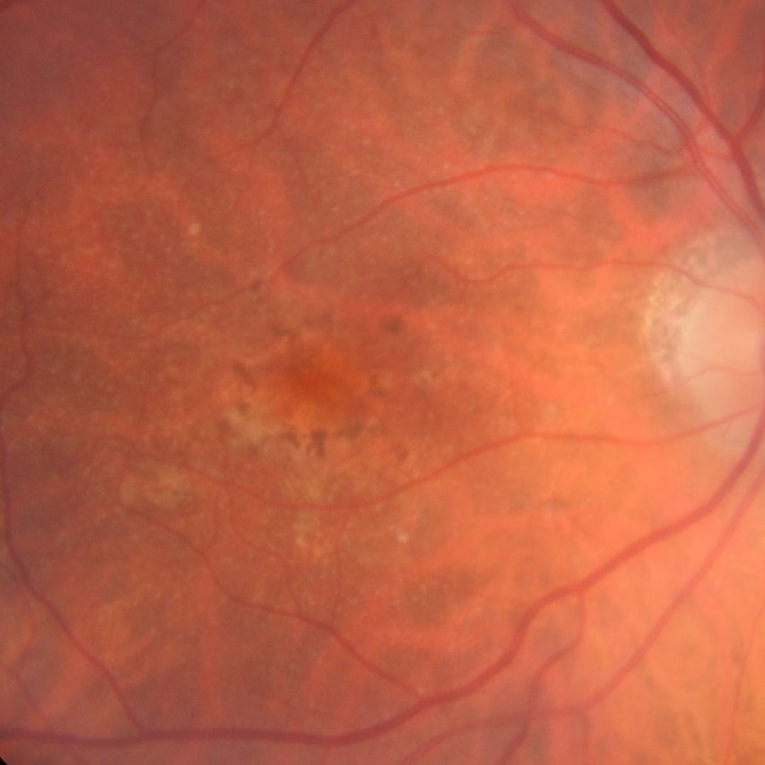
Drusen appear on colour fundus photography as yellow-white deposits and can vary significantly in size from small (less than 63µm) to large (125µm or more). Further information on estimating drusen size can be found at the bottom of this page.
It is important to note the location of drusen on OCT - underneath the RPE and above Bruch's membrane. Drusen have a rounded "dome-like" shape and exhibit homogenous internal reflectivity.
-
Reticular Pseudodrusen (RPD)
Reticular pseudodrusen (RPD), also termed subretinal drusenoid deposits, are distinct from other subtypes of drusen as they lie above the RPE. The presence of RPD in AMD is associated with a higher risk of progression to both forms of late AMD.
Colour fundus photography shows a yellowish interlacing or reticular pattern with a predilection for the superior macula.
Infrared imaging highlights the ‘target’ appearance of RPD. This refers to a central core with surrounding faint hyper autofluorescent halo and is a pattern also visible on fundus autofluorescence imaging.
OCT shows deposits above the RPE, in the subretinal space. They can be either flat or conical in shape.
-
Cuticular Drusen
Cuticular drusen are located between the RPE and Bruch's membrane, similar to the typical drusen. However, the multimodal imaging characteristics outlined below can help distinguish between the two variants.
Cuticular drusen are important to identify as they are associated with an increased risk of progression to geographic atrophy and choroidal neovascularisation. They may also present outside of the AMD spectrum.
They may present in 3 different phenotypes:
1. Densely populated in the macula or paramacular region
2. Scattered in the posterior fundus
3. Mixed with large drusenCuticular drusen appear as numerous coalescent small drusen on colour fundus photography. On infrared imaging, they can appear punctate hypo and hyper reflective, and may be less distinct than on colour fundus photography.
Fundus autofluorescence imaging typically shows punctate hypo autofluorescent dots with a hyperfluorescent surrounding.
OCT typically shows closely packed blunted triangles often described to exhibit a saw tooth appearance.
On fluorescein angiography, these drusen variants have been described as exhibiting a "starry sky" appearance.
Fundus photo (1), Red-free (2), Infra-red (3), Fundus autofluorescence (4) and Spectralis OCT (5)
More infoFundus photo (1), Red-free (2), Infra-red (3), Fundus autofluorescence (4) and Spectralis OCT (5)
More infoFundus photo (1), Red-free (2), Infra-red (3), Fundus autofluorescence (4) and Spectralis OCT (5)
More info -
Drusenoid PED
Drusenoid PED occur when drusen become confluent forming a pigment epithelial detachment. Its presence in AMD is associated with a higher risk of progression to the late stage.
On fundus photography, drusenoid PEDs are characterised by confluent drusen ½ disc diameter in size. They can have scalloped borders, be surrounded by soft drusen and may have a radiating pattern of hyperpigmentation on its surface.
OCT imaging typically shows a dome shaped RPE elevation with homogenous moderate internal reflectivity. Pigment migration can often be seen on its surface.
Fundus autofluorescence imaging can be variable but drusenoid PEDs most commonly show iso-autofluorescence or faint hyper-autofluorescence. Hyperpigmentation on the surface of the PED may lead to hyper-autofluorescence.
Fundus photo (1), Red-free (2), Infra-red (3), Fundus autofluorescence (4) and Spectralis OCT (5)
More infoFundus photo (1), Red-free (2), Infra-red (3), Fundus autofluorescence (4) and Spectralis OCT (5)
More infoFundus photo (1), Red-free (2), Infra-red (3), Fundus autofluorescence (4) and Spectralis OCT (5)
More info