
Overview
Bergmeister's papilla represents the remnants of the posterior hyaloid artery at the optic disc.
The hyaloid artery provides nutrition to the lens during embryonic development and then typically regresses as the foetus nears full maturation. If regression is only partial, a small glial tissue remnant remains and is termed Bergmeister's papilla. If a remnant remains attached to the posterior of the lens, it is called Mittendorf's dot.
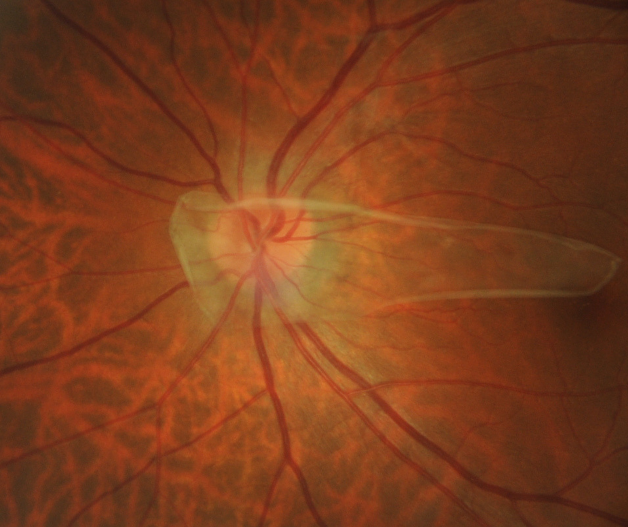
Fundus photography shows whitish collection of glial tissue anterior to the disc. The size and opacity of the tissue is dependent on the extent of remnant hyaloid artery.
OCT imaging shows a hyperreflective sheet of tissue located above the retinal plane and in the posterior vitreous space corresponding to the flap of glial tissue seen funduscopically.
Case Examples
Differential diagnosis
References
Jeon, H., Kim, J., & Kwon, S. (2019). OCT angiography of persistent hyaloid artery: a case report. BMC ophthalmology, 19(1), 141. https://doi.org/10.1186/s12886-019-1155-5
Sheth, J. U., Sharma, A., & Chakraborty, S. (2013). Persistent hyaloid artery with an aberrant peripheral retinal attachment: A unique presentation. Oman journal of ophthalmology, 6(1), 58–60.
Venkateswaran N, Moster SJ, Goldhagen BE. (2019) Bergmeister Papilla With Overlying Traction. JAMA Ophthalmol.137(9):e185915.