
Overview
The term megalopapillae is used to describe an abnormally large optic disc with a disc area greater than 2.5mm2.
Some features of megalopapillae may resemble glaucoma and this is an important differential to consider. Megalopapillae have large cup-to-disc ratios but should maintain an intact neuroretinal rim, RNFL thickness and visual field.
Megalopapillae may be bilateral with a normal cup configuration or unilateral with a superiorly displaced cup. Below are some examples of each.
For additional information on how to estimate disc size, please click on the link provided at the bottom of this page.
Case Examples
-
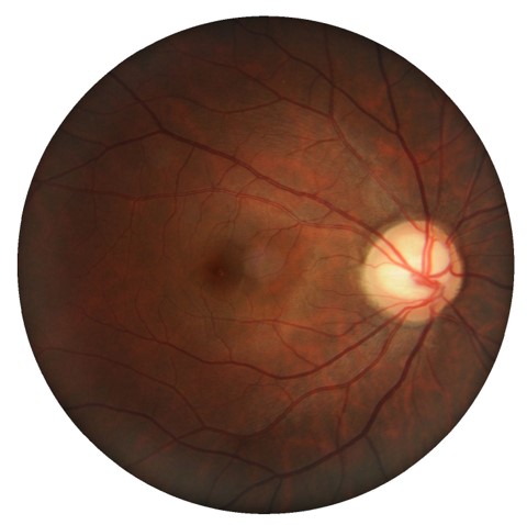
Case 1
A 34 year old Asian male with intraocular pressures of 16mmHg in each eye and central corneal thickness of 487µm (right eye) and 492µm (left eye). A 24-2 visual field test revealed enlarged blindspots in each eye, concordant with the enlarged discs seen on examination.
-
Case 2
A 48 year old Asian female with intraocular pressures of 13mmHg in each eye and central corneal thickness of 561µm (right eye), 563µm (left eye).
-
Case 3
A 58 year old Asian male with intraocular pressures of 18mmHg in each eye and central corneal thickness of 620µm (right eye) and 618µm (left eye.
Differential diagnosis
References
Ahuja Y, Traboulsi EI. (2010) Unilateral megalopapilla and contralateral optic nerve hypoplasia: a case report and review of the literature. J AAPOS. Feb;14(1):83-4.
Lee, H.S., Park, S.W. and Heo, H. (2015), Megalopapilla in children: a spectral domain optical coherence tomography analysis. Acta Ophthalmol, 93: e301-e305.