
- Overview
- Case Examples
- Differential diagnosis (optic disc coloboma)
- Differential Diagnosis (chorioretinal coloboma)
- References
Overview
An optic disc coloboma is characterised by a yellow white bowl-shaped excavation of the optic nerve, typically affecting the inferonasal part of the nerve, but in some cases involving the entire disc. Often associated with an optic disc coloboma are chorioretinal colobomas. These are typically found in the inferior retina and have a yellow-white appearance, often with hyperpigmented margins. OCT imaging shows excavation of the retinal and choroidal layers.
Optic nerve head colobomas arise from incomplete closure of the embryonic fissure prior to birth. This closure occurs when the embryo is approximately 10mm in size and at this stage, no retinal differentiation has occurred. Following this period of closure, the retina starts to develop distinct layers. The cells that lie over the top of a coloboma are actually still undifferentiated retinal cells - the cells that would otherwise in normal development develop into ganglion, amacrine, and Müller cells. These cells are referred to as the intercalary membrane.
Studies have shown that 23% to 42% of those with colobomas will eventually develop retinal detachment (Jesberg et al (1961) and Schepens et al (1983), frequently in the second decade of life. Choroidal neovascularisation can also occur at the edge of a coloboma with vessels growing in towards the intercalary membrane.
Case Examples
-
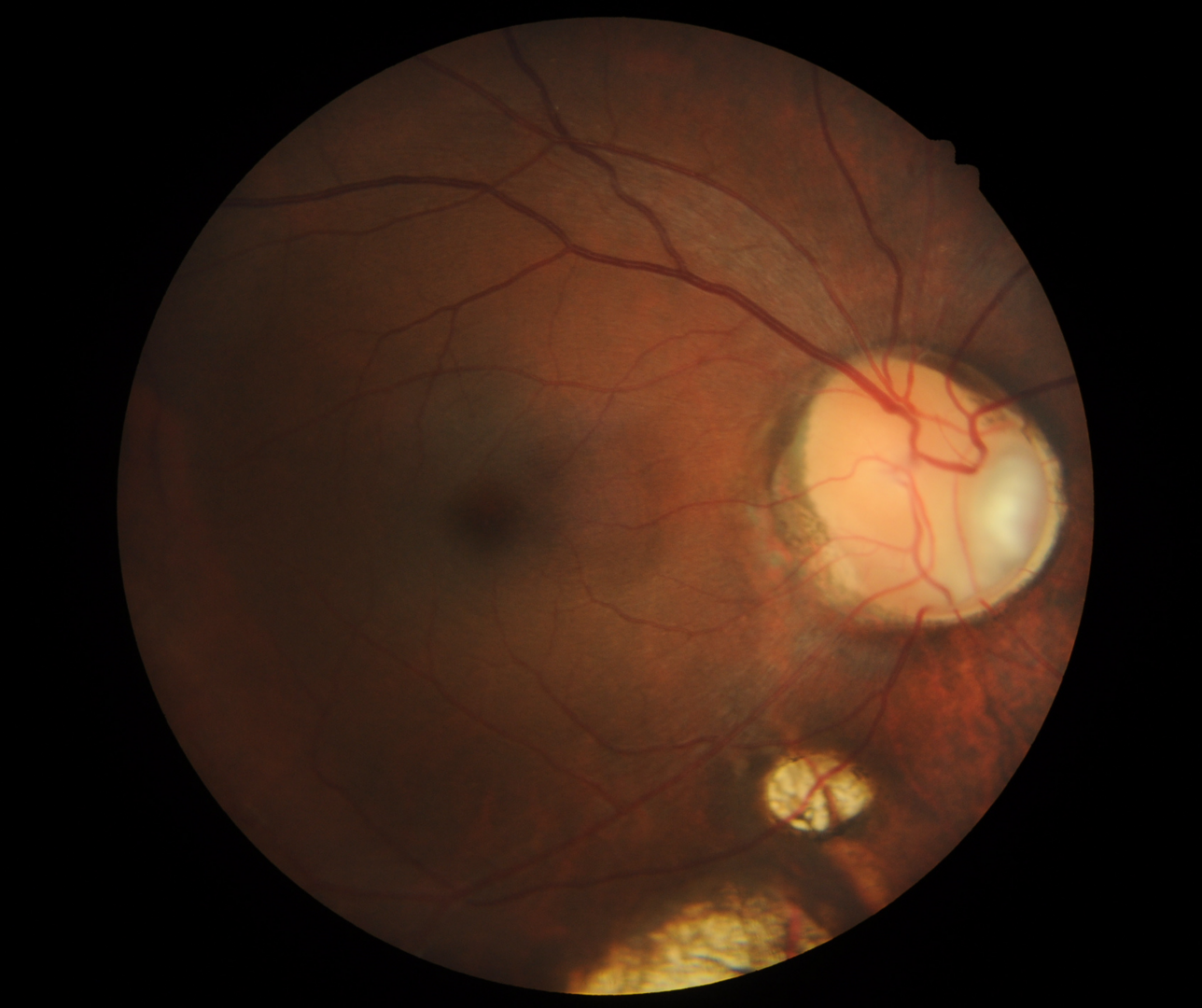
Case 1
A 43 year old Asian male with best corrected visual acuity in the left eye of 6/6 (20/20). Visual field testing revealed a superior absolute field defect corresponding to the areas of coloboma.
Fundus photograph (left eye)
More infoFundus photograph of the optic disc coloboma (left eye)
More infoOptomap (1), red separation (2) and green separation (3) images - left eye
More infoSpectralis OCT volume scan (left macular scan)
More infoB-scan ultrasound (left eye)
More infoSpectralis OCT line scan through the inferior chorioretinal coloboma
More info -
Case 2
A 43 year old Asian male with best corrected visual acuity in the right eye of 6/6 (20/20).
Fundus photograph (right eye)
More infoOptomap and green separation images (right eye)
More infoFundus auotfluorescence image (right eye)
More infoSpectralis OCT line scan through the right optic disc
More infoSpectralis OCT volume and line scan through chorioretinal coloboma (right eye)
More infoB scan ultrasound (right eye)
More info -
Case 3
An 8 year old female with best corrected visual acuity of 6/9 (20/30) in the right eye.
Fundus photograph and red free image (right eye)
More infoOptomap and green separation images (right eye)
More infoSpectralis OCT volume scan (right optic nerve head)
More infoSpectralis OCT line scan (right eye - inferior to the disc)
More infoCirrus RNFL analysis
More info24-2 SITA-Faster visual field
More info
Differential diagnosis (optic disc coloboma)
Differential Diagnosis (chorioretinal coloboma)
References
Kyoko Ohno-Matsui, Akito Hirakata, Makoto Inoue, Masahiro Akiba, Tatsuro Ishibashi; (2013) Evaluation of Congenital Optic Disc Pits and Optic Disc Colobomas by Swept-Source Optical Coherence Tomography. Invest. Ophthalmol. Vis. Sci;54(12):7769-7778.
Nakamura, K. M., Diehl, N. N., & Mohney, B. G. (2011). Incidence, ocular findings, and systemic associations of ocular coloboma: a population-based study. Archives of ophthalmology (Chicago, Ill. : 1960), 129(1), 69–74. https://doi.org/10.1001/archophthalmol.2010.320