
Overview
Optic nerve hypoplasia is an under-development of the optic nerve resulting in a reduced number of axons in the involved nerve. It may be bilateral or, more rarely unilateral (15-25% of cases). Supporting tissues and retinal vasculature develop normally. It may occur with in conjunction with microphthalmos, aniridia, coloboma, nystagmus, strabismus, facial anomalies, cranial abnormalities or occur independently of all these.
This condition is associated with a reduction in visual acuity that may range from light perception through to slightly sub-normal vision. Clinically, a very small optic nerve head is seen often surrounded by a ring of sclera.
OCT shows a thinning of the retinal nerve fibre layer (RNFL) and ganglion cell layer. The absence of a normal retinal nerve fibre layer can also be appreciated subjectively on red-free imaging which shows reduced reflectivity of the RNFL.
Case Examples
-
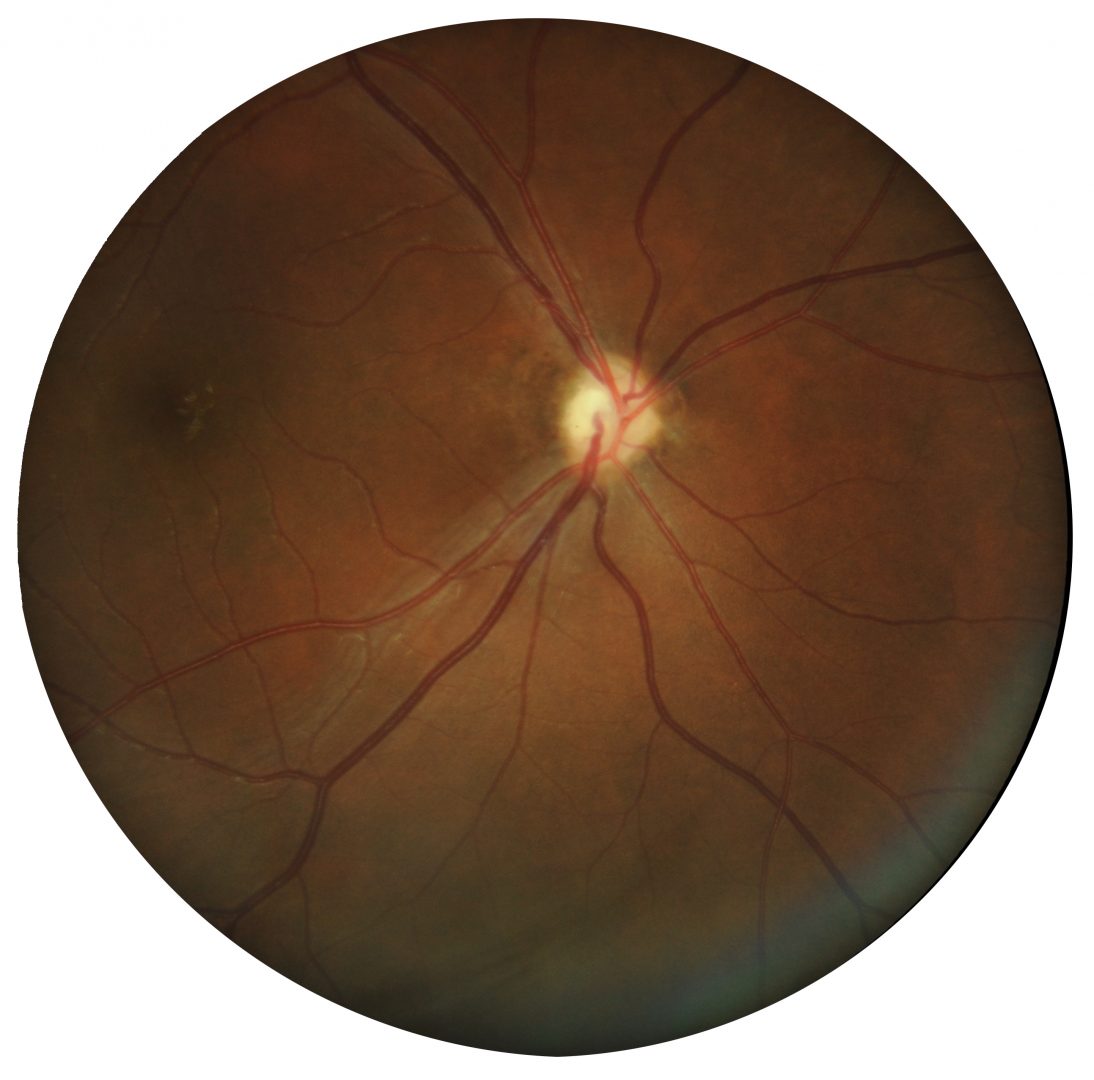
Case 1: Bilateral optic nerve hypoplasia
A 53 year old Caucasian male with best corrected visual acuity of 6/30 (20/100) in each eye. He has had poor vision since birth.
Fundus photographs (right and left eye)
More infoRed-free images (right and left eye)
More infoSpectralis OCT volume and line scans (right optic disc)
More infoSpectralis OCT volume and line scans (left optic disc)
More infoCirrus RNFL analysis
More infoCirrus ganglion cell analysis
More info30-2 SITA-standard visual field
More info -
Case 2: Unilateral optic nerve hypoplasia
A 28 year old Asian female with best corrected visual acuity of 6/24- (20/80-) in the right eye and 6/6 (20/20) in the left.
Differential diagnosis
References
Chen C, Yin J, Lewis RA, et al (2017) Genetic causes of optic nerve hypoplasia Journal of Medical Genetics 2017;54:441-449.
Garcia-Filion, P., & Borchert, M. (2013). Optic nerve hypoplasia syndrome: a review of the epidemiology and clinical associations. Current treatment options in neurology, 15(1), 78–89. https://doi.org/10.1007/s11940-012-0209-2