
Overview
Topless disc syndrome is a superior segmental optic disc hypoplasia thought to arise from an interruption in foetal development resulting in dysplasia of retinal neural tissue.
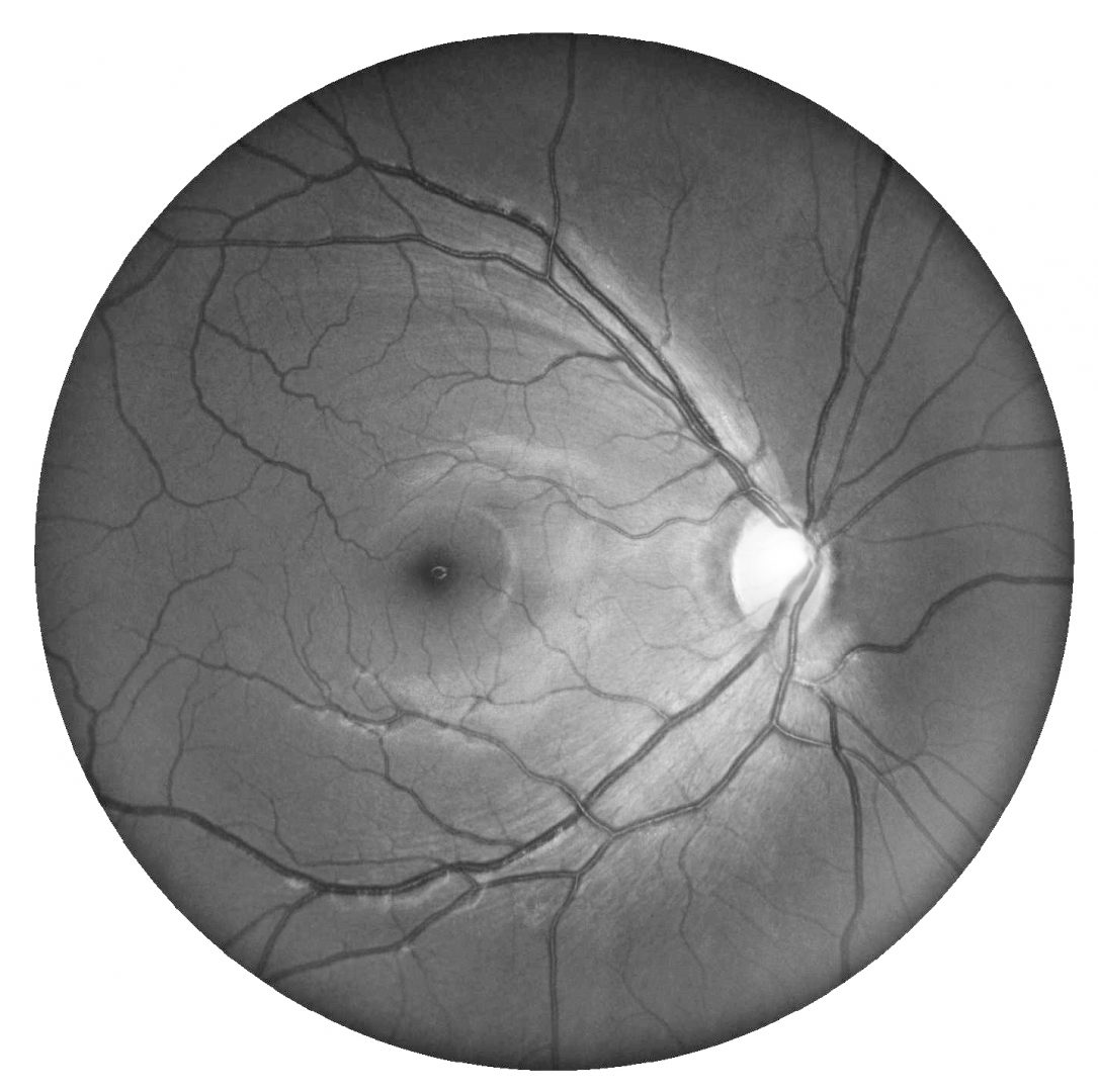
Funduscopically, a thinning of the RNFL and associated loss of the normal reflectivity is noted superiorly. Superior peripapillary atrophy and disc pallor with dense inferior altitudinal field defects are typically noted. Additionally, the superior branch of the central retinal artery enters the eye in a more superior location than is typical.
Topless disc syndrome has an association with maternal diabetes, however not all cases show this same association.
Case Examples
-
Case 1
A 39 year old asymptomatic Asian male with best corrected visual acuity of 6/6 (20/20) in both eyes.
Retinal photo and red-free image (right and left eye)
More infoDisc photo (1), red-free (2), OCT angiography superficial vascular plexus (3) and infra-red (4) images - right eye
More infoDisc photo (1), red-free (2), OCT angiography superficial vascular plexus (3) and infra-red (4) images - left eye
More infoCirrus Panomap analysis (right eye)
More infoCirrus Panomap analysis (left eye)
More infoGuided progression analysis (RNFL)
More infoGuided progression analysis (GCA)
More info30-2 SITA-Standard visual field
More info -
Case 2
A 30 year old asymptomatic Middle Eastern female with best corrected visual acuity of 6/6 (20/20) in the right eye.
Differential diagnosis
References
Shew, W. and Johnson, R.A. (2018), A case of topless disc syndrome (superior segmental optic hypoplasia). Clin Exp Optom, 101: 707-711
Sowka J, Vollmer L, Reynolds S. Superior segmental optic nerve hypoplasia: The topless disc syndrome. Optometry. 2008 Oct;79(10):576-80.