
Overview
Tilted disc syndrome is thought to arise from abnormal closure of the embryonic fissure and typically affects the inferonasal aspect of the retina.
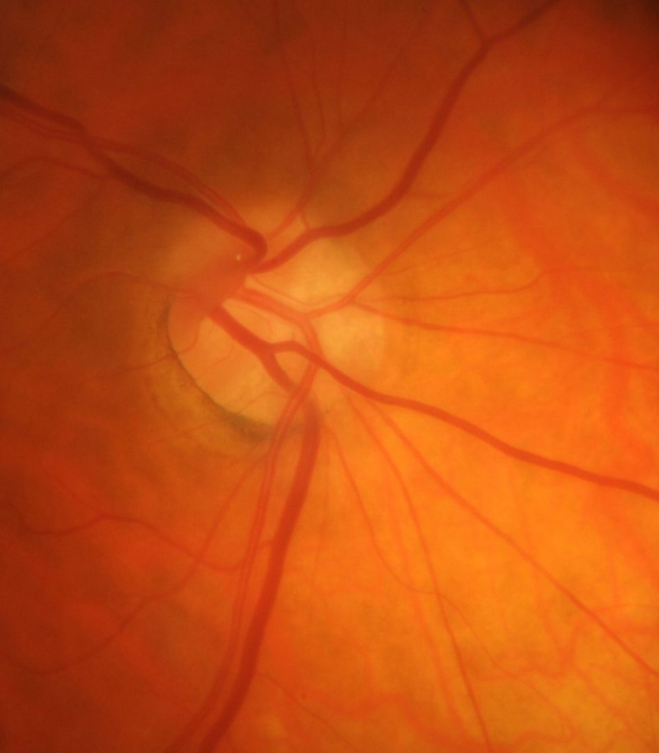
Features include an inferior or inferonasal tilted disc insertion with peripapillary atrophy and situs inversus of the emerging vasculature - a nasal diversion of the vessels as they emerge from the optic disc, followed by a sharp turn temporally. A posterior staphyloma can often be found in the inferonasal region of the fundus. The image here is of a tilted disc showing situs inversus in the right eye. Note that this syndrome is distinct from the tilted discs associated with progressive myopia.
The tilted nature of these discs can cause the nasal disc margin to appear indistinct due to a disparity between the maximum and minimum elevations of the surface of the disc. This can give rise to a pseudopapilloedema appearance.
OCT imaging through the optic nerve shows a sloped retinal plane with an elevation difference between the temporal and nasal aspect of the optic disc.
Temporal visual field defects can occur secondary to a refractive defocus (myopic) corresponding to the posteriorly bowed retina in the region of the posterior staphyloma. This visual field defect may be eliminated or reversed by using additional negative correction (e.g. -3.00D). This test is useful to differentiate between a temporal or bitemporal visual field defect caused by a lesion at the optic chiasm or by tilted disc syndrome.
Case Examples
-
Case 1
A 62 year old Caucasian female with best corrected visual acuity of 6/7.5 (20/25) in each eye.
-
Case 2
A 73 year old Caucasian male with best corrected visual acuities of 6/7.5- (20/25-) in both eyes. Mild posterior sub-capsular cataracts were noted.
Fundus photographs (right and left eye)
More infoStereoscopic image (right optic disc)
More infoStereoscopic image (left optic disc)
More infoSpectralis OCT line scan (left macula)
More infoCirrus RNFL analysis
More infoCirrus ganglion cell analysis
More info24-2 SITA standard visual field tests (normal correction and -3.00 overcorrection)
More infoB-scan ultrasound (left eye)
More info