
Overview
Consecutive optic atrophy is associated with diseases that affect the retina or its blood supply. This form of atrophy proceeds in an ascending manner whereby atrophy extends from a retinal cause through the axonal tissue towards the optic nerve. The underlying cause is usually obvious on fundus examination and causes include retinitis pigmentosa, vasculitis, retinal necrosis, retinitis or previous retinal photocoagulation.
Consecutive optic atrophy is typically a secondary diagnosis. The underlying condition is the primary diagnosis.
Case Examples
-
Case 1: Optic atrophy due to focal choroidal excavation
A 55 year old Asian male with best corrected visual acuity of 6/6 (20/20) in the right eye. The left eye is unremarkable, so this case will focus on the right eye only.
Retinal photo (1), red free image (2) and fundus autofluorescence images
More infoSpectralis OCT line scan through the focal choroidal excavation
More infoCirrus RNFL analysis
More infoCirrus ganglion cell analysis
More infoCirrus PanoMap analysis
More infoHumphrey 24-2 threshold visual field test
More info -
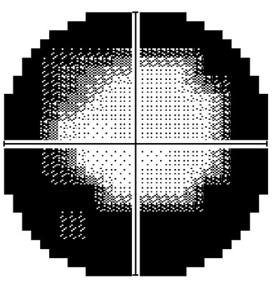
Case 2: Optic atrophy due to retinitis pigmentosa
A 67 year old Caucasian male, diagnosed with retinitis pigmentosa more than 30 years ago. His best corrected (central) visual acuity is 6/7.5 (20/25) in the right eye and 6/6- (20/20-) in the left.
Optomap widefield images (right and left eye)
More infoFundus autofluorescence images (right and left eye)
More infoSpectralis OCT volume and line scans (right macula)
More infoSpectralis OCT volume and line scans (left eye)
More infoCirrus RNFL analysis
More infoCirrus ganglion cell analysis
More infoHumphrey 10-2 threshold visual field analysis
More info