
Overview
Dominant optic atrophy (DOA) is an autosomal dominant disorder that usually presents in the first or second decade of life with slowly progressive vision loss. Additional features may include nystagmus and colour vision deficits. In the past, this condition was thought to be 2 separate conditions – a juvenile and an infantile form – however these are now regarded to be part of the same spectrum of disease.
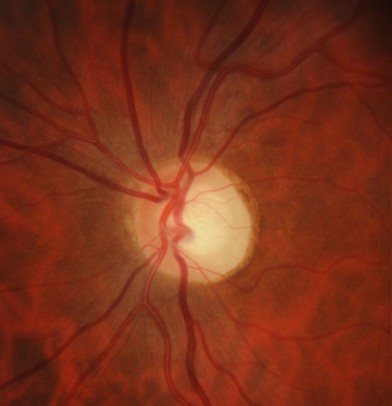
Funduscopic features include temporal disc pallor with a shallow 'saucerised' cupping of the neuroretinal rim.
OCT often shows marked temporal thinning of the RNFL with generalised thinning of the ganglion cell layer. Visual field testing typically shows a centrocecal defect.
Case Examples
-
Case 1
A 43 year old Caucasian female with a history of poor vision deteriorating since her teenage years. Her best corrected visual acuity was 6/19- (20/63-) in both eyes.
Optic disc photos and red free images (right and left)
More infoRed free images (right and left eyes)
More infoDesaturated D-15 colour vision
More infoSpectralis OCT macular line scans (right and left eye)
More infoCirrus RNFL analysis
More infoCirrus ganglion cell analysis
More infoCirrus macular thickness map
More info24-2 SITA-Standard visual field test
More info -
Case 2
A 45 year old Caucasian male with best corrected visual acuity of 6/7.5- (20/25-) in each eye. He reports difficulties driving at night.
Disc photos and red free images (right and left eye)
More infoRed free posterior pole images (right and left eye)
More infoSpectralis OCT macular line scans (right and left eye)
More infoCirrus PanoMap analysis (right eye)
More infoCirrus PanoMap analysis (left eye)
More info24-2 SITA-Standard visual field test
More info -
Case 3
A 45 year old Caucasian male with best corrected visual acuities of 6/30 (20/100) in the right eye and 6/24 (20/80) in the left eye. He has previously been diagnosed with DOA and has several family members also diagnosed with this condition.
Differential Diagnosis
References
Heidary G. (2014). Congenital optic nerve anomalies and hereditary optic neuropathies. Journal of pediatric genetics, 3(4), 271–280
Chun BY, Rizzo JF (2016). Dominant optic atrophy: updates on the pathophysiology and clinical manifestations of the optic atrophy 1 mutation. Curr Opin Ophthalmol. Nov;27(6):475-480.