
Overview
Primary optic atrophy occurs without preceding swelling of the optic disc. It can caused by lesions extending from the retina to the lateral geniculate nucleus. Causes include traumatic, toxic/nutritional, compressive or hereditary causes and retrobulbar optic neuritis.
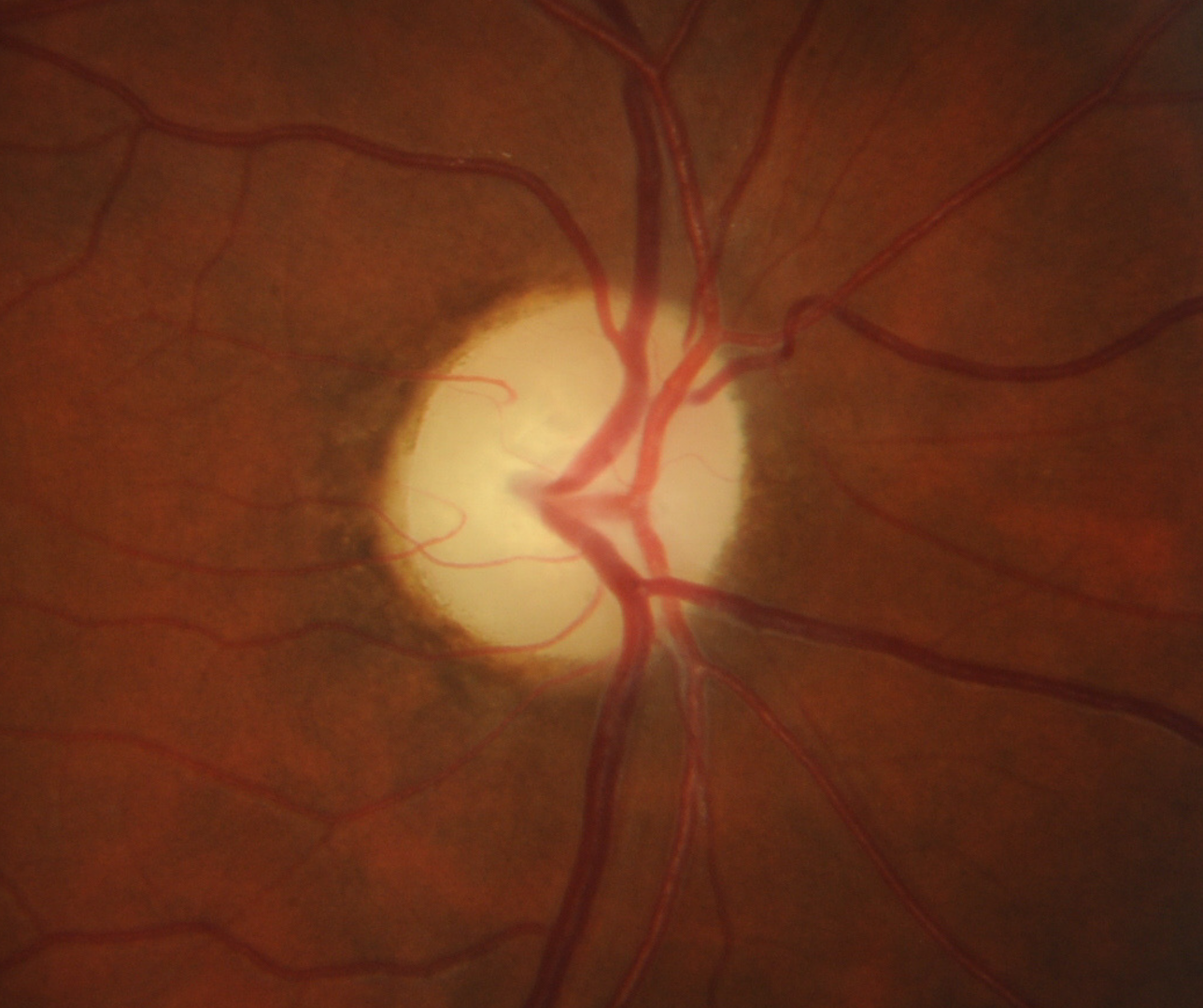
Funduscopic features include a chalky-white optic nerve appearance with very well defined disc margins. This is due to a loss of the usual RNFL that gives the margins of the optic disc a "softer" appearance. Also noteworthy is an eventual attenuation of the peripapillary blood vessels and a loss of the capillaries on the disc surface.
OCT analysis shows thinning of the retinal nerve fibre layer. Please note that the pattern of retinal nerve fibre layer loss is dependent on the cause and location of the lesion. Associated thinning of the retinal ganglion cell layer and visual field defects may also been seen.
Case Examples
-
Case 1: Traumatic optic atrophy
A 27 year old Cacuasian male with history of severe head trauma from falling off a roof 8 years ago. His visual acuity in the right eye is 6/12 (20/40) and the left 6/3 (20/10). The left eye is unremarkable, so this case will focus on the right eye.
-
Case 2: Optic atrophy due to optic nerve avulsion
A 40 year old Middle Eastern male with history of blunt trauma from a metal rod that impacted his right eye and lower lid 8 years previously. At the time he had a sudden loss of vision in the right eye which slowly resolved over several weeks but did not recover to the pre-injury level.
His best corrected visual acuity in the right eye is 6/12 (20/40).
Red cap test showed a 50% desaturation in the right eye.
Differential Diagnosis
References
Ahmad SS, Kanukollu VM. Optic Atrophy. [Updated 2020 Aug 11]. In: StatPearls [Internet]. Treasure Island (FL): StatPearls Publishing; 2020 Jan.
Osaguona V. B. (2016). Differential diagnoses of the pale/white/atrophic disc. Community eye health, 29(96), 71–74.