
Overview
Secondary optic atrophy refers to damage to retinal axons following an event of optic disc swelling. Common causes include papilloedema, optic neuritis and anterior ischaemic optic neuropathy.
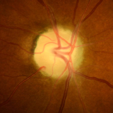
The degenerated RNFL is replaced at the optic disc by profuse glial cell proliferation which fills the cup. Funduscopic features include a grey-white disc appearance although the region of pallor may differ depending on the underlying cause. OCT typically shows RNFL and ganglion cell thinning concordant with the funduscopic region of disc pallor
Case Examples
-
Case 1: Previous papilloedema
A 46 year old Middle Eastern female with a history of papilloedema 9 years ago. Following investigations, no cause was found for the increased intracranial pressure. Her best corrected visual acuity is 6/9.5 in each eye (20/30).
-
Case 2: Previous papilloedema
A 43 year old Caucasian male with a history of meningitis causing increased intracranial hypertension at the age of 12. An MRI at the time was clear. His best corrected visual acuity is 6/4.8 in each eye (20/15).
References
Ahmad SS, Kanukollu VM. Optic Atrophy. [Updated 2020 Aug 11]. In: StatPearls [Internet]. Treasure Island (FL): StatPearls Publishing; 2020 Jan
Osaguona V. B. (2016). Differential diagnoses of the pale/white/atrophic disc. Community eye health, 29(96), 71–74.