
- Ocular Blood Supply
- Vascular Landmarks
- Vascular Disease Sub-Topics
- Vascular changes secondary to ocular disease
- References
Ocular Blood Supply
Arteries carry oxygenated blood from the heart to other organs, including the eye. To reach the eye, the blood travels via the common carotid artery to the internal carotid artery. This branches to form the ophthalmic artery which passes through the optic canal, infero-laterally to the optic nerve.
From here, several smaller arteries branch off the ophthalmic artery, including the short and long posterior ciliary arteries, the central retinal artery (CRA) and the lacrimal artery.
The CRA shares a common sheath with the central retinal vein (CRV) as it enters the eye. The narrowest lumen of the central retinal artery is where it pierces the dura mater of the optic nerve sheath passing through the lamina cribosa.
As the central retinal artery enters the eye, it divides into multiple branches to perfuse the inner layers of the retina. There is a small physiologically avascular area at the fovea approximately 1.5 degrees wide, known as the foveal avascular zone.
The outer retinal layers (including photoreceptors and the RPE) are perfused by the choroidal circulation which in turn is supplied by the long posterior ciliary arteries.
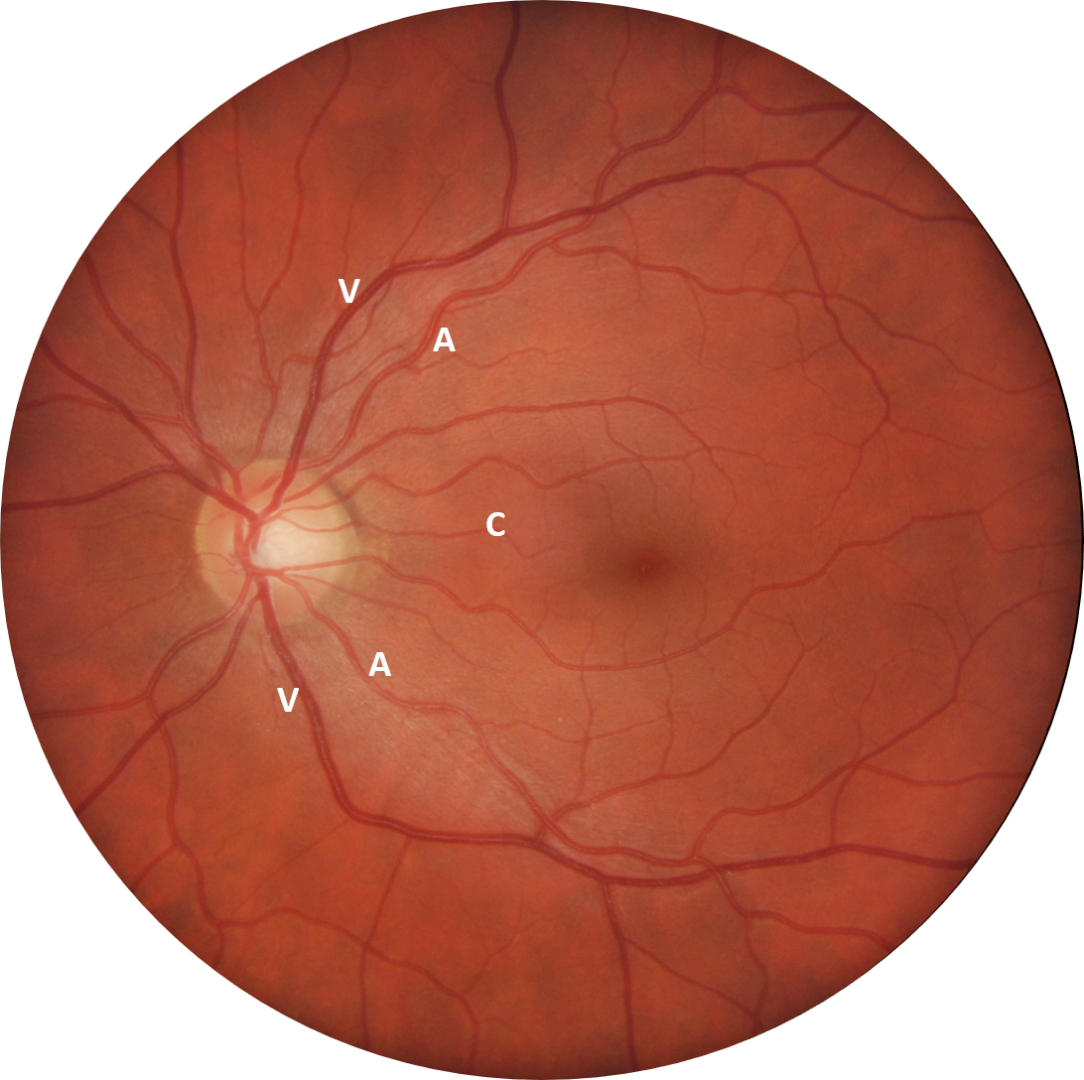
The short posterior ciliary arteries perfuse the optic disc via the circle of Zinn-Haller. Additionally, in approximately 50% of people, the short posterior ciliary artery also has an additional, anomalous branch that supplies the area of retina between the optic disc and macula - a cilioretinal artery (marked "C" in this image).
Retinal arteries (marked A in the image) are narrower than retinal veins (marked V), while the veins are darker in colour than the arteries as they are carrying de-oxygenated blood. At the point where the vessels cross the optic disc, an artery is typically 2/3 the width of a vein.
The headings below link to a discussion of various vascular anomalies of the posterior eye.
Vascular Landmarks
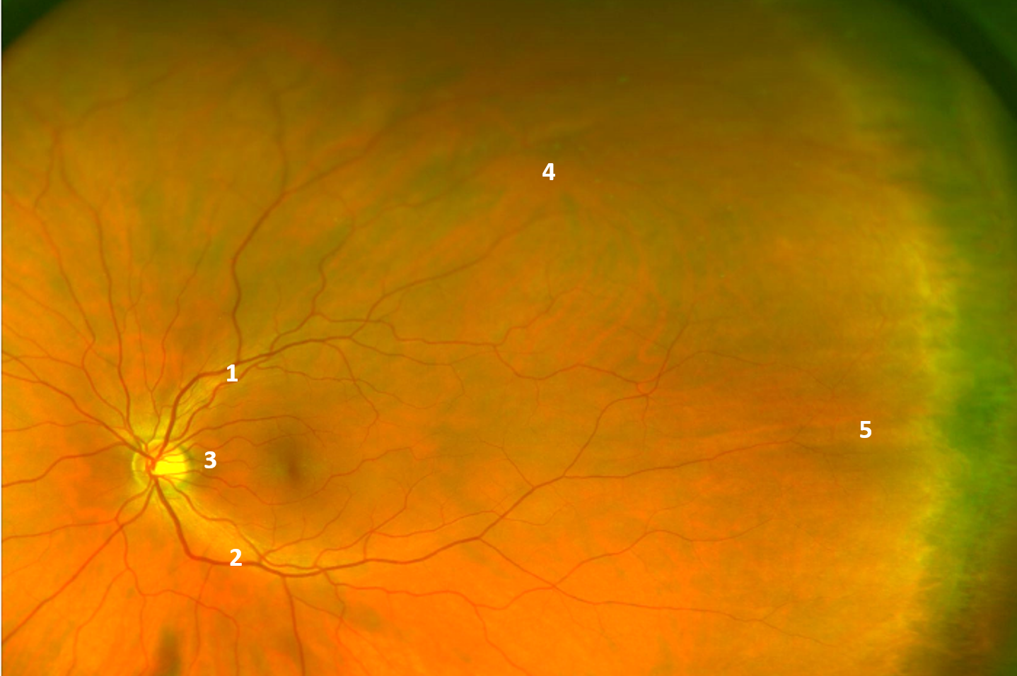
The widefield image shown here illustrates the key vascular features of the retina. The superior vascular arcades (1) and inferior vascular arcades (2) emerge from the optic disc, running temporally in an arcuate shape. These vessels are branches of the central retinal artery which divide further as you move away from the optic nerve.
The cilioretinal artery (3) is visible on this image also.
The vortex vein ampullae (4) are an important anatomical landmark as they define the location of the equator. These may vary in number from 4 to 8 in each eye, however most people will have 4 or 5. A vortex vein ampulla may be described as the convergence of multiple large choroidal vessels into a single large ampullar trunk.
The long posterior ciliary nerve (5), while not vascular in nature, may be seen on peripheral examination and it is helpful to be able to identify this also. Adjacent to the long posterior ciliary nerve is the dark band of the ciliary body. The point at which this joins the retina is termed the ora serrata, and it typically has a serrated appearance.
Vascular Disease Sub-Topics
Vascular changes secondary to ocular disease
References
Snell, R. Lemp, M. (2013) Clinical Anatomy of the Eye 2nd Ed. Pub Wiley-Blackwell