
Coats disease
Coats disease is characterised by retinal telangiectasias and exudation and is typically diagnosed between ages 8 and 16 years. Most cases are unilateral (95%) and the condition occurs most frequently in males (75%).
Visual acuity may be reduced by several mechanisms, including cystoid macular oedema, exudative retinal detachment and/or neovascular glaucoma.
Symptoms and/or signs may include reduced vision, strabismus and/or leukocoria however some patients are asymptomatic at diagnosis. This reflects the range of clinical presentations possible. Mild cases may show only a few affected (telangiectatic) vessels in the peripheral retina, while advanced cases may show retinal detachment, corneal oedema and/or iris neovascularisation.
OCT imaging typically shows subtle intraretinal edema or subretinal fluid. OCTA and fluorescein angiography show vascular abnormalities (telangiectatic vessels) with bulb-like aneurysms. Capillary dropout and areas of retinal non-perfusion may also be seen.
A staging system for Coats’ disease was developed by Jerry Shields et al. Very briefly, stage 1 is characterised by retinal telangiectasia, stage 2 by the development of extra-foveal and foveal exudation, stage 3 by the development of exudative retinal detachment, stage 4 by the onset of secondary glaucoma and stage 5 is end-stage disease. For further detail, please refer to the Shields et al (2001) paper listed in the references at the bottom of this page.
Case Example
-
Case 1
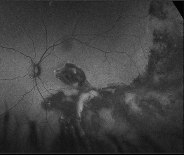
A 29 year old Caucasian female with recent onset flashes and floaters, diagnosed with eye disease in her left eye at the age of 13. Her visual acuity in the left eye was counting fingers at 50cm.
Fundus photograph (left eye)
More infoRed-free image (left eye)
More infoOptomap (1), green separation (2) and fundus autofluorescence imaging (3)
More infoCirrus OCT Angiography (left eye)
More infoSpectralis OCT volume and line scan (left eye)
More infoPeripheral OCT line scan (left eye, infero-temporal retina)
More info
Differential diagnosis
References
Shields, J, Shields, CL, Honavar,SG, Demirci,H, Cater,J. (2001) Classification and management of Coats disease: the 2000 Proctor Lecture,American Journal of Ophthalmology, Volume 131, Issue 5, Pages 572-583,
Shields CL, Udyaver S, Dalvin LA, et al (2020) Visual acuity outcomes in Coats disease by classification stage in 160 patients. British Journal of Ophthalmology 104:422-431.
Yousef, YA, Rimawi, AH, Nazzal, RM, Qaroot, AF, AlAref, AH, Mohammad, M et al. (2020). Coats’ disease: characteristics, management, outcome, and scleral external drainage with anterior chamber maintainer for stage 3b disease, Medicine: April 2020 - Volume 99 - Issue 16 - p e19623