
Overview
A retinal macroaneurysm is an acquired dilation (aneurysm) of a retinal arteriole, believed to arise from a combination of hypertension and arteriosclerosis. Retinal signs may be quite variable, making diagnosis challenging. To that end we have provided several examples below of retinal macroaneurysm.
Retinal macroaneurysms are typically found at either arteriovenous crossings or arterial bifurcation, most commonly within the first 3 birfucations of the central retinal artery. The macroaneurysm presents as a rounded dilation within an artery that may or may not show spontaneous pulsation. Exudates may be present, usually in a circinate pattern surrounding the aneurysm. Ruptured aneuryms result in subretinal, intraretinal, preretinal and/or vitreal haemorrhage.
Most retinal macroaneurysms are asymptomatic, however if the macula area is affected or rupture results in a vitreous haemorrhage, patients will present with an acute or gradual vision loss.
Retinal macroaneurysms will often spontaneously resolve over a period of several months however the strong association of these lesions with hypertension necessitates communication with the patient's general practitioner.
Case Examples
-
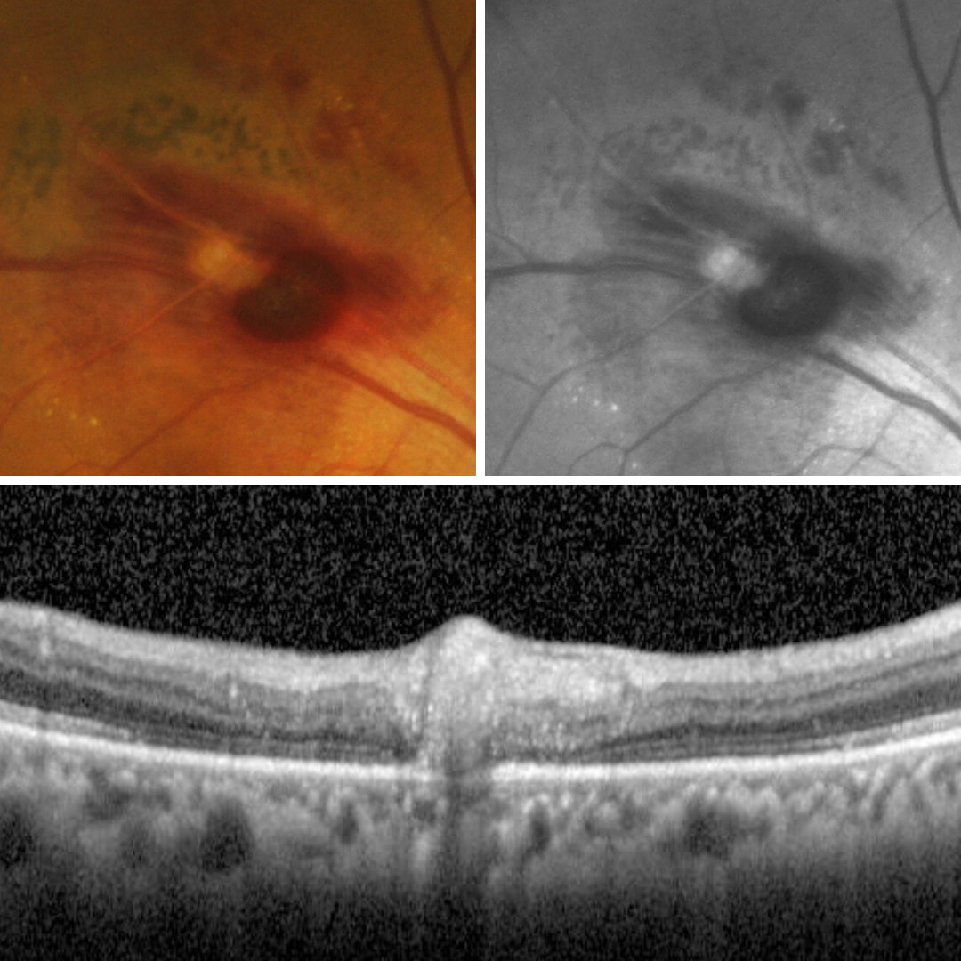
Case 1: Small macroaneurysm
A 76-year-old Caucasian male with a history of systemic hypertension for which he is taking medication. His best corrected visual acuity is 6/6 (20/20) in the left eye. This case will focus on the left eye only.
-
Case 2: Ruptured macroaneurysm
A 76-year-old Asian male with type 2 diabetes (diagnosed 20 years previous), hypertension and hypercholesterolaemia, all of which are treated with medication. He has nuclear sclerosis in the left eye and best corrected visual acuity of 6/15 (20/50) in this eye.
-
Case 3: Resolving macroaneurysm
A 79-year-old Asian male with treated hypertension.
-
Case 4: Thrombosed retinal macroaneurysm
A 75-year-old Caucasian female with grade 3 nuclear sclerosis in the right eye and best corrected visual acuity of 6/12 (20/40) in this eye.
-
Case 5: Retinal macroaneurysm
A 66 year-old Caucasian male who smokes more than 20 cigarettes a day and has done so for many years. He is on medications for heart arrhythmia and hypertension and takes an anticoagulant.
Differential Diagnosis
References
Moosavi, R., Fong, K. & Chopdar, A. (2006) Retinal artery macroaneurysms: clinical and fluorescein angiographic features in 34 patients. Eye 20, 1011–1020.
Speilburg, A. M., & Klemencic, S. A. (2014). Ruptured retinal arterial macroaneurysm: diagnosis and management. Journal of optometry, 7(3), 131–137.