
Overview
Hypertension (high blood pressure) is a chronic medical condition in which the blood pressure in the arteries is elevated. It carries an increased risk of cardiovascular disease, including coronary artery disease, ischaemic heart disease, stroke, renal failure and more.
According to guidelines published by the Heart Foundation, normal blood pressure ranges from 120-129 systolic, and 80-84 diastolic. Above this, the following categories apply:
High normal: Systolic 130-139; Diastolic 85-90
Grade 1 (mild) hypertension: Systolic 140-159; Diastolic 91-99
Grade 2 (moderate) hypertension: Systolic 160-179; Diastolic 100-109
Grade 3 (severe) hypertension: Systolic greater than or equal to 180; Diastolic greater than or equal to 110.
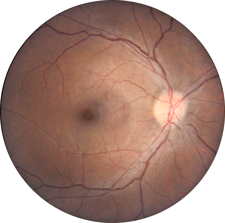
Chronic hypertension causes changes to the vasculature of the body, and these may be visualised in the retina. It is important to remember this as hypertensive changes noted on retinal examination are likely to reflect non-visible changes elsewhere in the body. For this reason, communication with a patient's GP when hypertensive changes are noted is highly advisable.
When classifying hypertensive retinopathy, this Atlas will refer to the Mitchell-Wong Simplified Hypertensive Retinopathy Grading Scale, as detailed below:
MILD: Generalised arteriolar narrowing, focal arteriolar narrowing, arteriovenous nicking (nipping), copper wiring (opacity of arteriolar wall) or a combination of these signs
MODERATE: Retinal haemorrhages (blot-shaped, dot-shaped or flame-shaped), microaneurysm, cotton wool spot, hard exudate or a combination of these signs
MALIGNANT: Signs of moderate retinopathy plus swelling of the optic disc
Following are some examples of the pathological changes listed in the grading scale outlined above.
Arteriovenous Changes
-
Arteriolar narrowing
Hypertension can cause generalised or focal arteriolar narrowing.
-
Copper wiring
Copper wiring refers to an increased light reflex of the retinal arterioles due to atherosclerotic changes that increase vessel wall thickness. The vessel lumen appears orange-coloured rather than red.
-
Silver wiring
Silver wiring is a more advanced form of copper wiring where the light reflex of the retinal arterioles is whiter (more silver-white) than in copper wiring.
-
AV crossing changes
AV nipping (AV nicking) refers to the indentation or narrowing of a vein caused by an overlying arteriosclerotic artery. Either side of the artery, the vein shows a reduced diameter.
Hypertension may also result in other arteriovenous crossing changes such as the deflection of a vein by an arteriole through to a 90 degree banking of a retinal vein. Some examples of arteriovenous changes in hypertensive retinopathy are given here.
Retinal Signs
-
Flame haemorrhage
A flame haemorrhage is located in the retinal nerve fibre layer, and this locale is responsible for the characteristic linear shape as the blood accumulates between the retinal nerve fibres.
-
Cotton wool spot
Cotton wool spots arise due to the interruption of axoplasmic flow, typically due to ischaemia. Cotton wool spots present on OCT as a focal thickening of the retinal nerve fibre layer.
-
Hard Exudates
Hard exudates are associated with retinal or macular oedema and present as focal yellow lesions in the retina. The hard exudates appear as hyper-reflective foci on OCT, primarily located in the outer nuclear and outer plexiform layers. The term macular star refers to the presence of numerous hard exudates, radiating out from the macula radially in all directions.
Optic Nerve signs
-
Optic nerve head oedema
Optic nerve head oedema (papilloedema) occurs in cases of malignant hypertension and is a medical emergency. Signs of papilloedema include disc indistinct margins, the presence of Paton folds, "rainbow rings" on OCT RNFL analysis, V-contour on OCT and symptoms including nausea and/or headaches. All of these signs and symptoms are covered in greater depth in this Atlas (please use the "papilloedema" link at the bottom of the page to learn more)
Sub-Topics
References
Grosso A, Veglio F, Porta M, et al (2005) Hypertensive retinopathy revisited: some answers, more questions British Journal of Ophthalmology 89:1646-1654.
Tsukikawa, M., & Stacey, A. W. (2020). A Review of Hypertensive Retinopathy and Chorioretinopathy. Clinical optometry, 12, 67–73.
Wong,TY, McIntosh,R (2005) Hypertensive retinopathy signs as risk indicators of cardiovascular morbidity and mortality, British Medical Bulletin, Volume 73-74, Issue 1 Pages 57–70,
Wong,TY, Mitchell,P (2004) Hypertensive Retinopathy. N Engl J Med 351:2310-2317