
Overview
Moderate hypertensive retinopathy is characterised by retinal changes that include retinal haemorrhages (blot-shaped, dot-shaped or flame-shaped), microaneurysms, cotton wool spots, hard exudates or a combination of these signs. These signs indicate a breakdown of vascular autoregulatory mechanisms and are an acute sign of hypertensive damage.
Several of the signs and symptoms of moderate hypertensive retinopathy are also associated with diabetic retinopathy and it can be challenging to determine the the primary cause of retinal changes if the patient's medical history is positive for both hypertension and diabetes.
Case Examples
-
Case 1
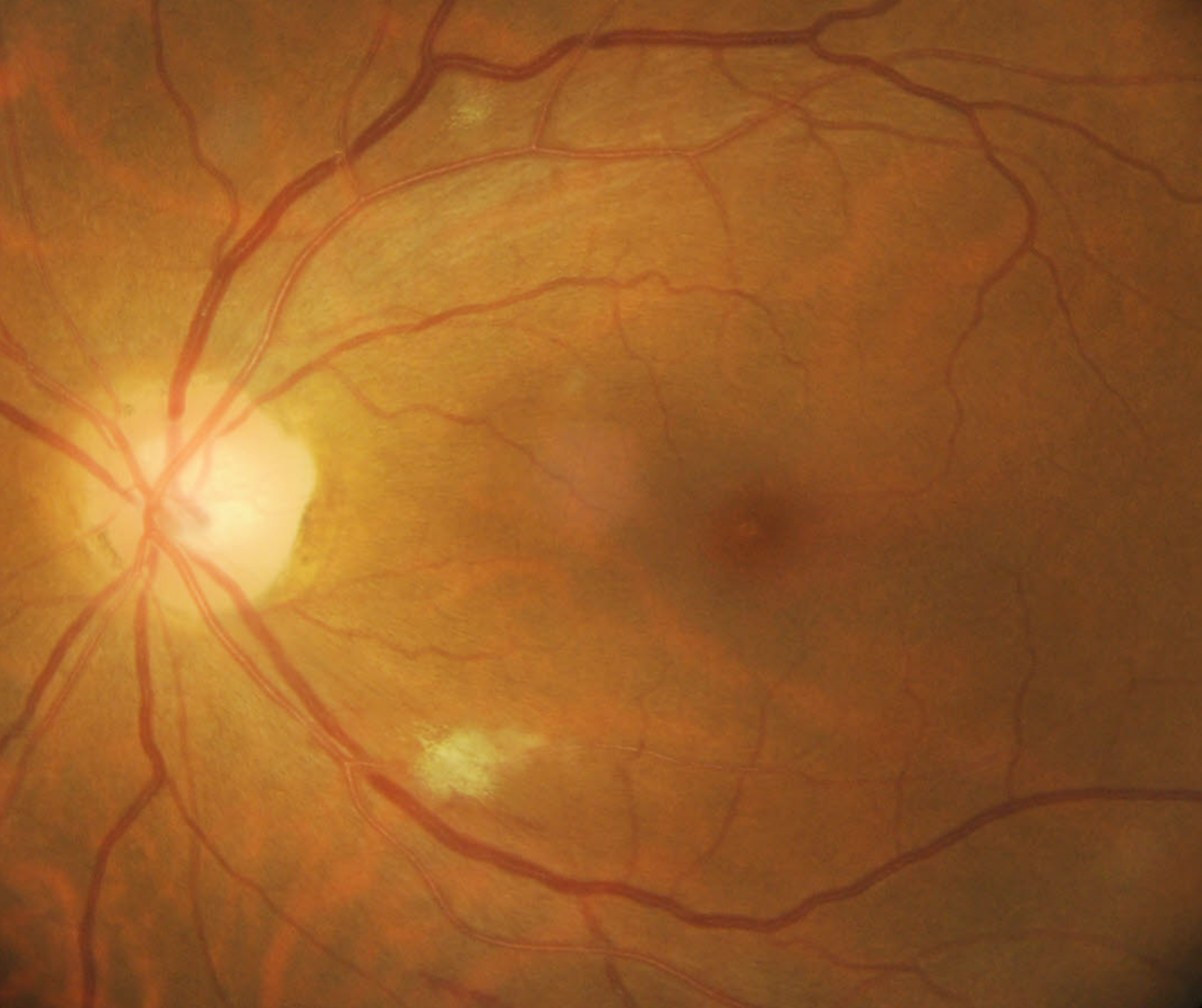
A 49-year-old Indian male who has a history of hospitalisation for an acute increase in blood pressure 4 months earlier. His blood pressure is now treated with medication. His in-office blood pressure was measured at 171/110 mmHg. His best corrected visual acuity was 6/6 (20/20) in each eye.
Given his history of acute hypertension, in combination with the clinical appearance of the retina, his GP was contacted and he was referred for a prompt ophthalmology consult.
-
Case 2
A 56-year-old Caucasian female with no reported significant medical history. Her best corrected visual acuity is 6/6 (20/20) in the right eye and 6/7.5 (20/25) in the left. In-office blood pressure measurement was 167/94 mmHg.
A report was sent to this patient's GP regarding the elevated blood pressure measurement and associated retinopathy and the patient was encouraged to make an appointment promptly.
Differential Diagnosis
References
Wong,TY, Mitchell,P (2004) Hypertensive Retinopathy. N Engl J Med 351:2310-2317