
Overview
A branch retinal artery occlusion (BRAO) occurs when an embolus becomes lodged in a retinal artery, completely blocking the flow of oxygenated blood to the area of retina supplied by that artery. Whilst a CRAO affects the entire retina, a BRAO affects a segment of it only.
Vision in BRAO may be variable depending on the location of the occlusion, but most commonly acuity is better than 6/12.
As with other types of retinal arterial occlusion, retinal ischaemia ensues; the retina takes on a whitish, opaque appearance in the acute stages and OCT may show an associated thickening of the RNFL in the ischaemic area.
BRAO is also associated with arterial attenuation and "cattle trucking" - a term that refers to segmentation of the blood downstream to the occlusion.
In the chronic stage significant thinning of the inner retina can be noted in the area impacted by the BRAO. A permanent visual field defect may be associated with this thinning.
Retinal neovascularisation can occur following a BRAO, however the incidence of this is relatively low.
Case Examples
-
Case 1: Acute BRAO
A 77-year-old Caucasian female with best corrected visual acuity in the right eye of 6/12 (20/40). She takes medications for arthritis, heart disease and hypercholesterolaemia. Her left eye is unremarkable so this case will focus on her right eye.
Fundus photograph (right eye)
More infoRed-free image (right eye)
More infoOptomap image and green separation image (right eye)
More infoSpectralis OCT line scan through ischaemic retina (right eye)
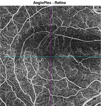
More infoOCT Angiography (right eye, 6x6mm, superficial vascular plexus)
More infoCirrus OCT ganglion cell analysis (3 month follow-up)
More info -
Case 2: Chronic BRAO
A 55-year-old Middle Eastern male with type 2 diabetes, diagnosed 5 years previously. His best corrected visual acuity in the right eye of 6/7.5 (20/25).
Differential Diagnosis
References
Chen, C., Lee, A. (2008) Management of acute central retinal artery occlusion. Nat Rev Neurol 4, 376–383.
Fallico, M., Lotery, A.J., Longo, A. et al. (2020) Risk of acute stroke in patients with retinal artery occlusion: a systematic review and meta-analysis. Eye 34, 683–689
Woo, S., Lip, G. & Lip, P. (2016) Associations of retinal artery occlusion and retinal vein occlusion to mortality, stroke, and myocardial infarction: a systematic review. Eye 30, 1031–1038.
Varma, D., Cugati, S., Lee, A. et al. (2013) A review of central retinal artery occlusion: clinical presentation and management. Eye 27, 688–697.