
Overview
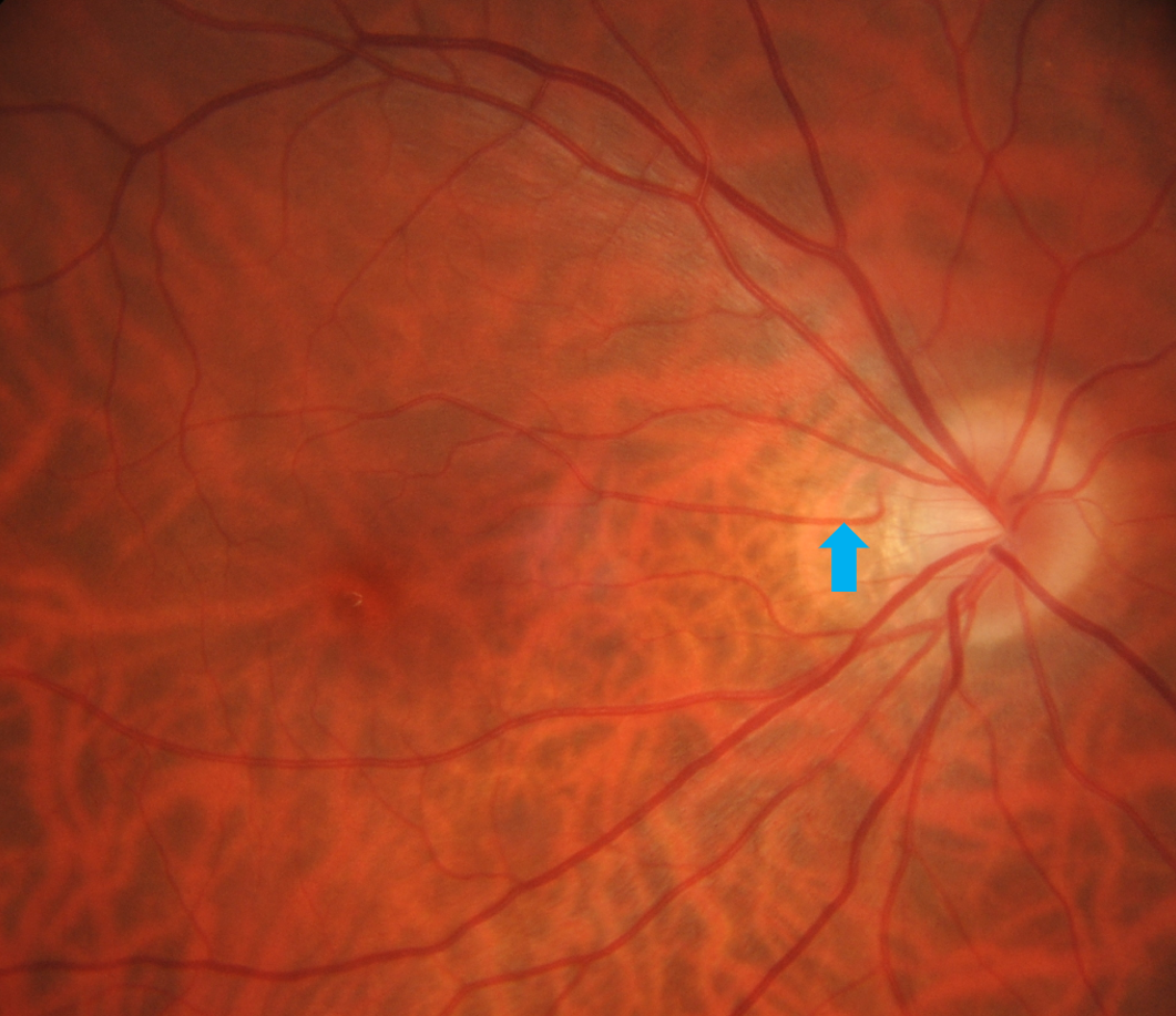
Up to 50% of the population have an additional anomalous branch of the short posterior ciliary artery or peripapillary choroid that exits the disc temporally, radiating out towards the macula. An example of a cilioretinal artery is shown in the image (indicated by the blue arrow).
As with other retinal arteries and arterioles, the cilioretinal artery may become occluded by embolism. In the acute phase, a cilioretinal artery occlusion results in an area of white, ischaemic retina in the papillomacular area and OCT shows oedema of the inner retinal layers.
In the chronic stages, as with other types of arterial occlusion, atrophy/thinning of the inner retina may be noted on OCT. Disc pallor may also be noted.
Case Example
-
Case 1: Chronic cilioretinal artery occlusion
A 50-year-old Caucasian male with hypercholesterolaemia and best corrected visual acuity of 6/7.5 (20/25) in his right eye.
Differential Diagnosis
References
Messner LV, Newman TL, Bartlett M, Conto JE. Cilioretinal artery occlusion with central retinal vein occlusion (1999) Optometry and Vision Science : Official Publication of the American Academy of Optometry. Nov;76(11):741-746.
Schneider, M., Molnar, A., Angeli, O., Szabo, D., Bernath, F., Hajdu, D., Gombocz, E., Mate, B., Jiling, B., Nagy, B.V., Nagy, Z.Z., Peto, T. and Papp, A. (2021), Prevalence of Cilioretinal Arteries: A systematic review and a prospective cross‐sectional observational study. Acta Ophthalmol.
Stoffelns BM. Isolated cilioretinal artery occlusion - clinical findings and outcome in 31 cases. (2012) Klinische Monatsblatter fur Augenheilkunde. Apr;229(4):338-342.