
Overview
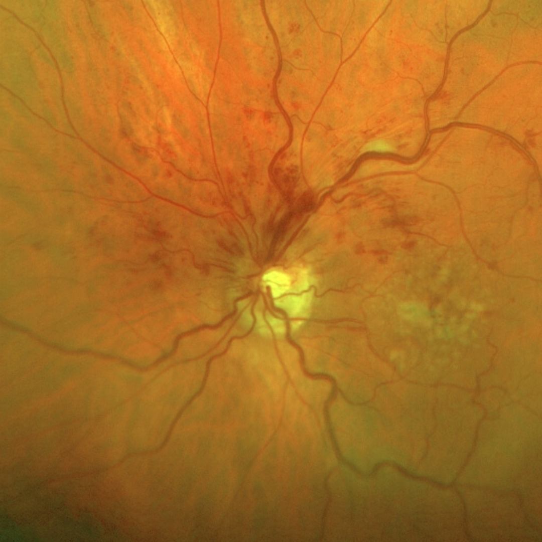
A HRVO refers to an occlusion of either the superior or inferior branch of the central retinal artery. In this condition, half of the retina shows the classic signs of retinal venous occlusion, including dilated tortuous veins and widespread retinal haemorrhages. The opposing half of the retina is unaffected.
As with other types of venous occlusion, HRVO may also be associated with retinal or macular oedema, cotton wool spots and the development of either collateral vessels or neovascularisation.
Case examples
-
Case 1: Superior HRVO
A 70 year old Caucasian male who has noted blurring in his left eye for the last month. He has a history of deep vein thrombosis. Testing with an Amsler grid showed distortion inferior to fixation in the left eye and his best corrected visual acuity in this eye is 6/12- (20/40-).
Differential Diagnosis
References
Jia Li, Yannis M. Paulus, Yuanlu Shuai, Wangyi Fang, Qinghuai Liu, and Songtao Yuan (2017) New Developments in the Classification, Pathogenesis, Risk Factors, Natural History, and Treatment of Branch Retinal Vein Occlusion. Journal of Ophthalmology Volume 2017 |Article ID 4936924.
Khayat, Survey Ophth 2018. Major Review: Ischemic retinal vein occlusion: characterising the more severe spectrum of retinal vein occlusion
Patel, A., Nguyen, C., & Lu, S. (2016). Central Retinal Vein Occlusion: A Review of Current Evidence-based Treatment Options. Middle East African journal of ophthalmology, 23(1), 44–48.
Woo SCY , Eye 2016. Associations of RAO and RVO to mortality, stroke and myocardial infarction: a systematic review