
Overview
Pigmented paravenous chorioretinal atrophy is characterised by pigmentation and associated chorioretinal atrophy along the retinal veins.
This is an uncommon disorder of unknown aetiology, although an inherited, degenerative, or inflammatory cause has been proposed.
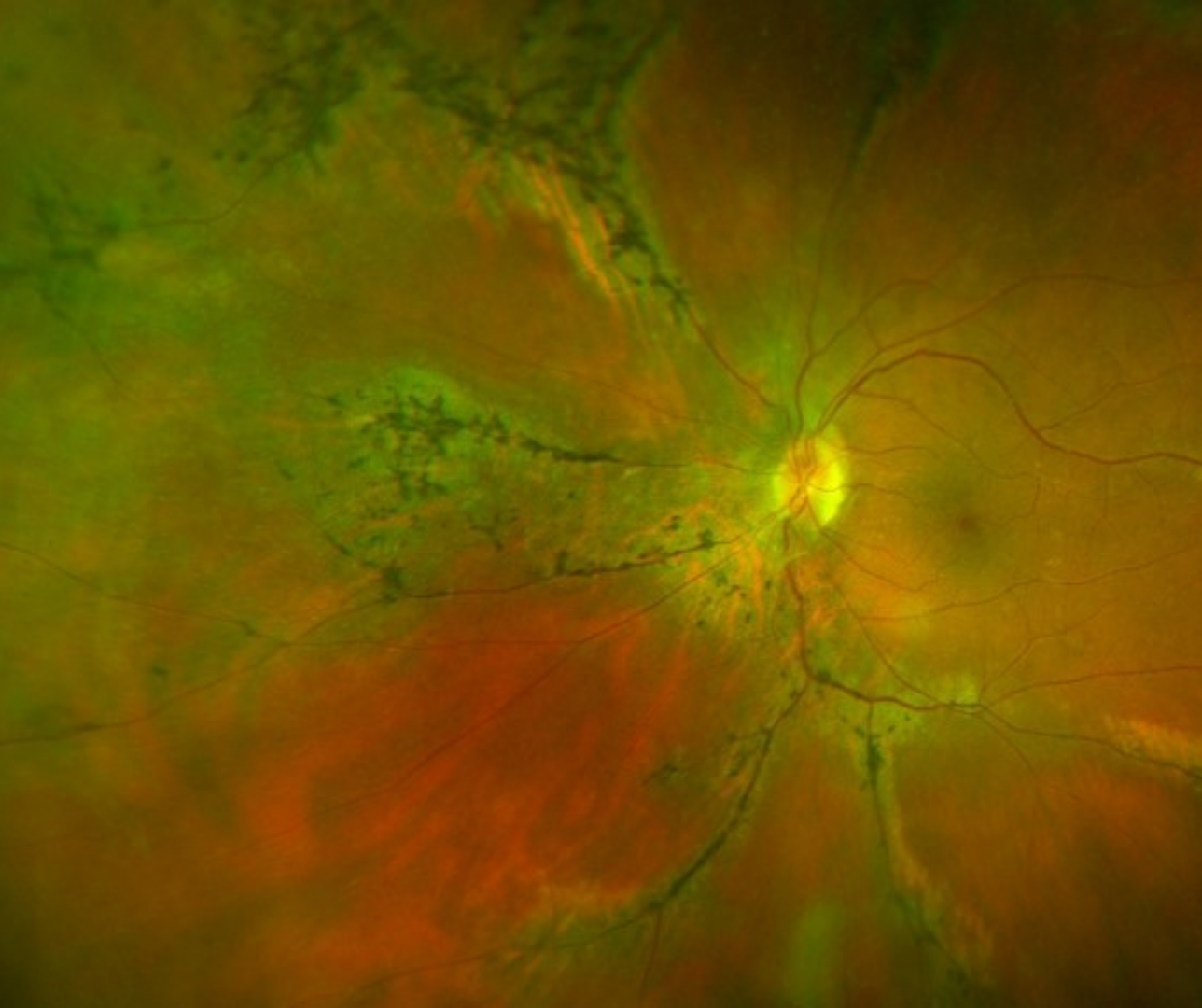
Funduscopically, an accumulation of pigment is noted, distributed along the retinal veins. Areas of chorioretinal atrophy are noted adjacent to the vascular pigmentary changes.
OCT typically shows thinning of the outer retinal layers with increased backscattering and disorganisation of the RPE-choriocapillaris complex. Areas of hyper-pigmentation in the retina correspond to hyper-reflective intra-retinal plaques with posterior shadowing (pigment migration).
OCTA shows reduced perfusion in the choriocapillaris in the areas of chorioretinal atrophy, with relative sparing of the retinal capillary plexus.
Fundus autofluorescence typically shows a linear hypo-autofluorescence along the large retinal veins, surrounded by an area of hyper-autofluorescence.
Case Examples
-
Case 1: Pigmented paravenous chorioretinal atrophy (paravenous type)
A 65-year-old Caucasian female with best-corrected visual acuity of 6/6+ (20/20+) in each eye.
-
Case 2: Pigmented paravenous chorioretinal atrophy
A 36 year old, asymptomatic Middle Eastern female with best corrected visual acuity of 6/6 (20/20) in each eye.
Optomap widefield and green separation images (right eye)
More infoOptomap widefield and green separation images (left eye)
More infoFundus autofluocescence images
More infoCirrus OCT line scan (right inferior vascular arcade)
More infoCirrus OCT line scan (left supero-temopral retina)
More info
Differential Diagnosis
References
Lee, EK, Lee, SY, Oh, BL, Yoon, CK, Park, UC, Yu, HG (2021) Pigmented Paravenous Chorioretinal Atrophy: Clinical Spectrum and Multimodal Imaging Characteristics, American Journal of Ophthalmology, Volume 224, Pages 120-132.
Shona, O. Islam, F. Robson, AG; Webster, AR er al. (2019) Pigmented paravenous chorioretinal atrophy. Retina: March 2019 - Volume 39 - Issue 3 - p 514-529