
Overview
Ocular conditions affecting the retinal blood supply and inner retina can lead to the development of retinal neovascularisation. The process of retinal neovascularisation involves the growth of new capillaries from the existing retinal vasculature in response to hypoxia and the subsequent release of vascular endothelial growth factor (VEGF).
Retinal neovascularisation is broadly described as either neovascularisation of the disc (NVD) or neovascularisation elsewhere (NVE).
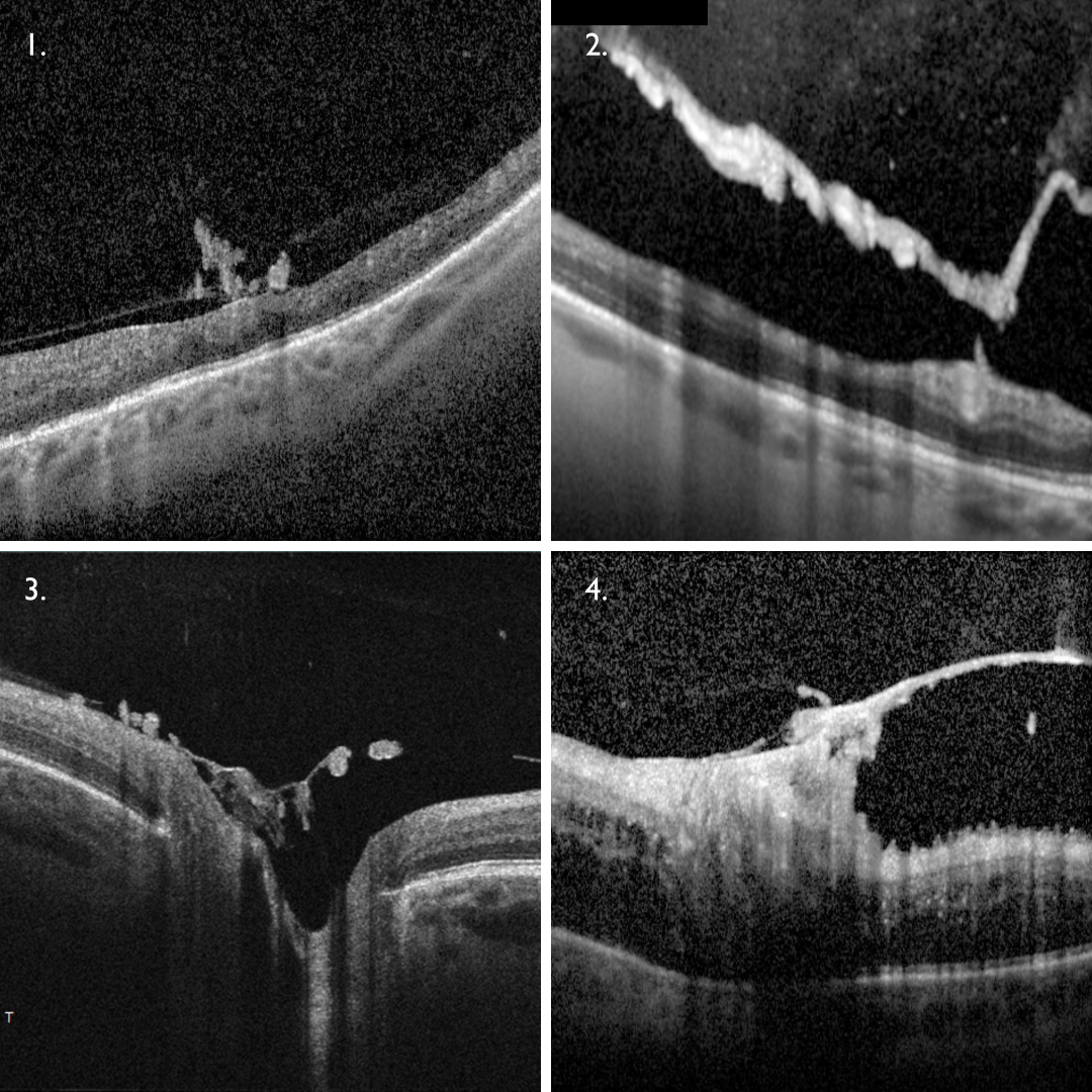
NVE presents on OCT as either hyper-reflective homogenous loops on the retinal surface that show posterior shadowing (1), or areas of hyper-reflectivity on the posterior hyaloid interface (2). OCT-A can confirm the presence of fine, irregular vessels within the vitreoretinal interface.
NVD appears as hyperreflective material sitting on optic disc surface and/or protruding into the vitreous (3). More advanced NVD can appear as a fibrovascular tissue overlying the disc with associated traction (4). OCT-A can confirm the presence of an irregular network of vessels above the optic disc surface.
Conditions giving rise to retinal neovascularisation are listed below with links to the relevant chapters within this resource.
Associated Conditions
References
Lee, P. Wang, CC. Adamis, AP. (1998) Ocular Neovascularization: An Epidemiologic Review,
Survey of Ophthalmology. Volume 43, Issue 3, pp 245-269.
Vaz-Pereira, S., Morais-Sarmento, T. Esteves Marques, R. (2020) Optical coherence tomography features of neovascularization in proliferative diabetic retinopathy: a systematic review. Int J Retin Vitr 6, 26.