
Overview
A Roth spot is a flame haemorrhage with a characteristic white centre. These haemorrhages are associated with a variety of systemic disease, most notably with bacterial endocarditis. Other associated conditions include leukemia, anaemia, hypertensive retinopathy, pre-eclampsia, diabetic retinopathy, human immunodificiency virus (HIV), pre-eclampsia, shaken baby syndrome and anoxia.
Identification of a Roth spot should prompt follow-up with a medical practitioner to investigations related to possible systemic disease.
Roth spots are the result of retinal capillary rupture and intraretinal hemorrhage. The white centre of the lesion is composed of fibrin that has formed a fibrin-platelet "plug" at the site of capillary rupture.
Case Examples
-
Case 1
A 70 year old Middle Eastern male with type 2 diabetes, hypertension and hypercholesterolaemia. Best corrected visual acuity 6/6 in each eye.
-
Case 2
A 59 year olf Middle Eastern female who was diagnosed with type 2 diabetes 15 years previously. Her most recent HbA1c was 7.4% and her best corrected visual acuity is 6/6 in each eye.
-
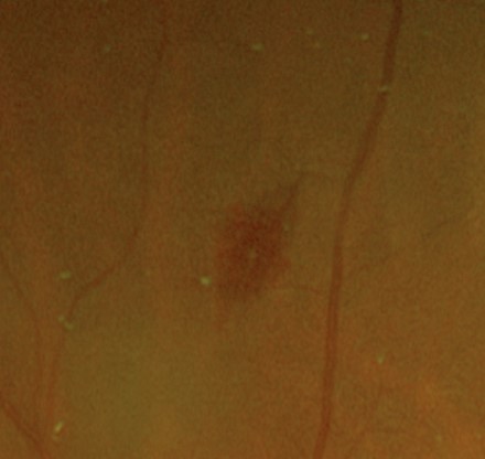
Case 3
A 17 year old Asian male with a genetic haematological condition that causes anaemia. His best corrected visual acuity is 6/6 (20/20) in each eye.