
Overview
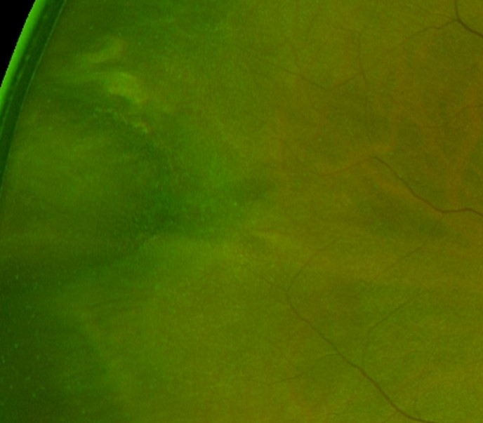
An oral bay is formed by the joining of two or more dentate processes at the ora serrata. These dentate processes may join (completely enclosed) or have a gap remaining (partially enclosed oral bay).
Oral bays may be confused with a retinal hole on examination however oral bays do not increase the risk of retinal detachment.
Oral pearls are drusen-like deposits located between the RPE and Bruch’s membrane. The deposits are white and glistening on examination and may be found within an oral bay or adjacent to the dentate processes.
Case Examples
-
Case 1
An asymptomatic 31-year-old Caucasian male with best corrected visual acuity of 6/6 (20/20) in the left eye.
-
Case 2
An asymptomatic 35-year-old Caucasian female with best corrected visual acuity of 6/6 (20/20) in her right eye.
-
Case 3
An asymptomatic 38-year-old Caucasian female with best corrected visual acuity of 6/6 (20/20) in the right eye.
Differential diagnosis
References
Campagnoli T.R., Smiddy W.E. (2016) Peripheral Retinal Abnormalities. In: Medina C., Townsend J., Singh A. (eds) Manual of Retinal Diseases. Springer, Cham
Salmon, S., Bowling, B. Kanski, J. (2019) Kanski’s clinical ophthalmology – A systematic approach. 9th edition.