
- Overview
- Case Examples
- Retinal breaks & lattice
- Examples of retinal breaks
- Differential Diagnosis
- References
Overview
Lattice degeneration is characterised clinically by a linear or round, typically circumferentially oriented area of retinal thinning with abnormal vitreoretinal adhesion and associated traction at the edges. It is typically located in the peripheral retina (between the equator and the ora serrata) and is bilateral in 34%-48% of cases. Rarely, lattice lesions may present posterior to the equator is which case they are typically oriented radially.
Lattice degeneration may present with fine white interlacing lines caused by sclerosed vessels (a similar appearance to lattice) internally, underlying areas of hyper-pigmentation and/or with small yellow-white particles at the lesion margins. The retina is thinned across the surface of the lesion and this may lead to atrophic holes within the lesion.
Retinal thinning and vitreoretinal traction can be appreciated with peripheral OCT imaging. Some presentations of lattice show focal areas of increased thinning and full thickness atrophic hole formation.
OCT imaging may demonstrate adherence of the vitreous at the margins of the lesion and a clear pocket of liquid vitreous over the central part of the lesion. This manifests on OCT as a U-shaped appearance.
Case Examples
-
Case 1: Bilateral lattice
A 61-year-old asymptomatic Caucasian male with best corrected visual acuity of 6/6 (20/20) in each eye.
Optomap (1) red separation (2) and green separation (3) images - Right eye
More infoOptomap (1) red separation (2) and green separation (3) images - left eye
More infoMagnified view of lattice degeneration in each eye and fundus autofluorescence images (right -1,3 and left 2,4)
More infoSpectralis OCT through lattice (left temporal retina)
More info -
Case 2: Lattice degeneration in a lightly pigmented fundus
A 33-year-old Caucasian female with best corrected visual acuity of 6/6 (20/20) in her left eye.
-
Case 3: Heavily pigmented lattice
A 57-year-old asymptomatic Caucasian male with best corrected visual acuity of 6/6 (20/20) in his left eye.
Fundus photograph of the area of lattice (superotemporal retina, left eye)
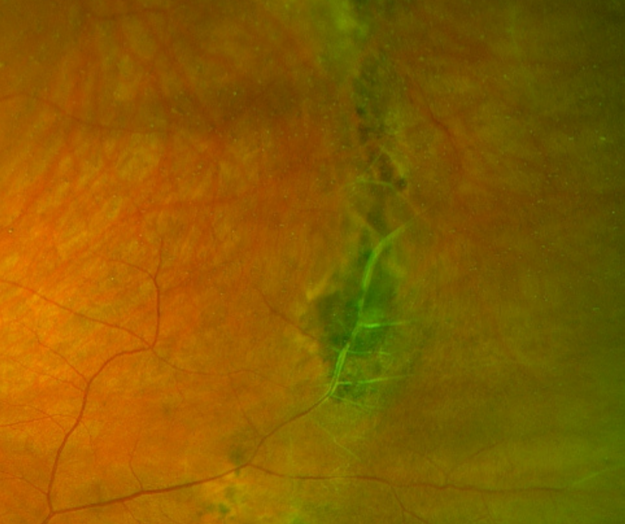
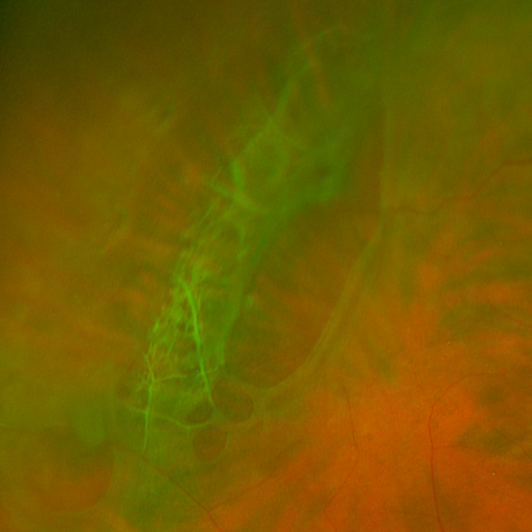
More infoOptomap (1) red separation (2) and green separation (3) images - left superotemporal retina
More infoSpectralis OCT vertical line scan taken through the area of lattice (left superotemporal periphery)
More infoSpectralis OCT line scan taken through the edge of an area of lattice
More infoSpectralis OCT line scan through the area of lattice degeneration
More info
Retinal breaks & lattice
Vitreoretinal traction at the edges of lattice lesions can cause subclinical retinal detachments and sometimes lead to retinal breaks at the posterior edge of the lattice (the edge of the lesion closest to the posterior pole) following a posterior vitreous detachment. With ongoing vitreoretinal traction, retinal breaks associated with lattice degeneration may continue on to rhegmatogenous retinal detachment (RRD).
Approximately 20% to 30% of patients with RRD have lattice degeneration, so the presence of lattice is considered a risk factor for retinal detachment. The risk of a retinal tear or detachment is increased if perivascular or radial lattice is present.
Atrophic holes internal to the lattice may cause a localised sub-clinical detachment but rarely progress beyond that. These holes may be single or multiple and are found in 16-24% of eyes with lattice degeneration.
Examples of retinal breaks
-
Case 1: Lattice with internal atrophic holes
A 30-year-old asymptomatic Caucasian female with moderate myopia and best corrected visual acuity of 6/6 (20/20) in the right eye.
-
Case 2: Retinal tear associated with lattice
A 60-year-old asymptomatic Caucasian male with best corrected visual acuity of 6/6 (20/20) in the right eye.
-
Case 3: Horseshoe tear associated with lattice
An asymptomatic 71-year-old Caucasian female with best corrected visual acuity of 6/6 (20/20) in the left eye.
-
Case 4: Retinal detachment associated with lattice
An asymptomatic 72-year-old Caucasian female with best corrected visual acuity of 6/6 (20/20) in the right eye.
-
Case 5: Bilateral holes associated with lattice
A 24-year-old asymptomatic Asian male with best corrected visual acuity of 6/6 (20/20) in each eye.
Differential Diagnosis
References
Flaxel, C. Adelman, RA, Bailey, ST, Lim, JI et al. (2020) Posterior Vitreous Detachment, Retinal Breaks, and Lattice Degeneration Preferred Practice Pattern. Ophthlamology. Volume 127, Issue 1, pp146-181.
Lewis, H (2003) Peripheral retinal degenerations and the risk of retinal detachment, American Journal of Ophthalmology, Volume 136, Issue 1, pages 155-160,
Manjunath, V., Taha, M., Fujimoto, J. G., & Duker, J. S. (2011). Posterior lattice degeneration characterized by spectral domain optical coherence tomography. Retina (Philadelphia, Pa.), 31(3), 492–496.