
Overview
A meridional fold is a radially oriented, thickened area of retinal tissue, found in the peripheral retina perpendicular to the ora serrata. It appears grey-white in colour and has a prevalence of 26%.
A meridional fold is typically aligned with dentate processes and cystoid degeneration is often found within the fold.
Rarely, a meridonial fold may develop a small retinal hole in the retina adjacent to the posterior limit of the fold (the closest end to the posterior pole). This hole forms due to vitreo-retinal traction at the tip of the lesion.
Case Examples
-
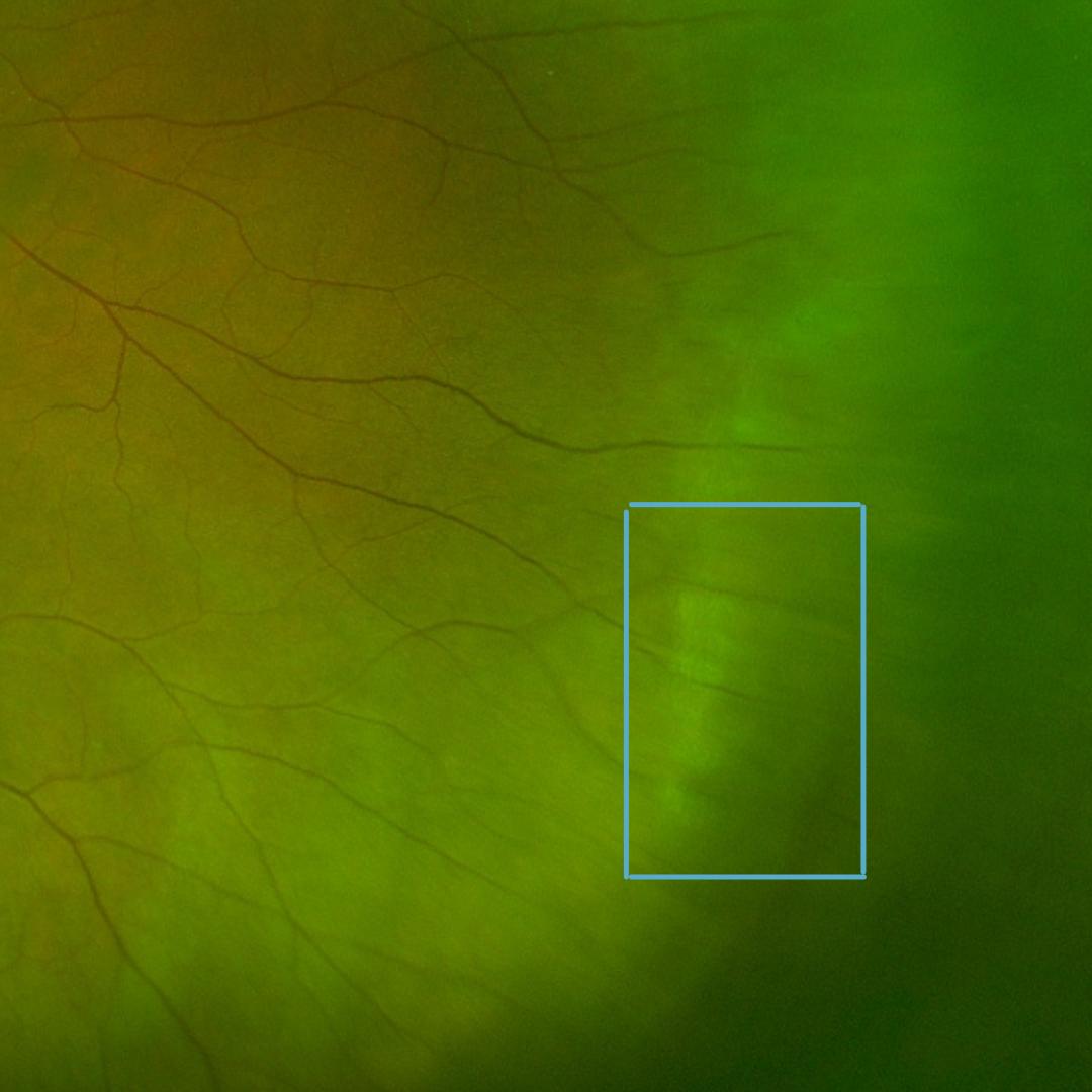
Case 1
A 29 year old asymptomatic Asian female with best corrected acuity of 6/6 (20/20).
References
Campagnoli T.R., Smiddy W.E. (2016) Peripheral Retinal Abnormalities. In: Medina C., Townsend J., Singh A. (eds) Manual of Retinal Diseases. Springer, Cham. https://doi-org.wwwproxy1.library.unsw.edu.au/10.1007/978-3-319-20460-4_49
Salmon, S., Bowling, B. Kanski, J. (2019) Kanski’s clinical ophthalmology – A systematic approach. 9th edition.