
Overview
There is debate in the literature as to whether snail-track degeneration is actually an early-stage presentation of lattice degeneration or its own entity. Regardless, this condition does have several clinical features in common with lattice degeneration.
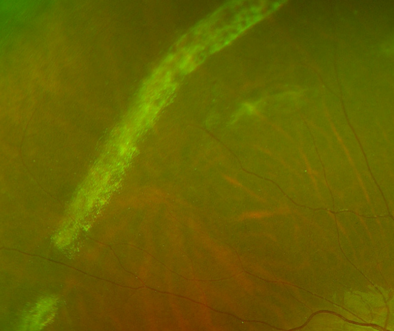
Snailtrack degeneration is characterised by glistening yellow-white linear bands in the peripheral retina. These bands resemble lattice degeneration, however don’t have the typical network of fine “lattice-like” lines found in lattice degeneration. Snailtrack degeneration is not associated with underlying hyperpigmentation.
Similar to lattice degeneration however, snailtrack degeneration shows abnormal vitreoretinal adhesions, vitreous liquefaction (lacuna) overlying the degeneration and retinal thinning. It may be associated with atrophic holes and horseshoe tears and (rarely) retinal detachment.
Snailtrack degeneration is most commonly found in the inferotemporal peripheral retina but can occur in other areas.
Case Examples
-
Case 1
A 27-year-old asymptomatic Middle Eastern male with best corrected visual acuity of 6/6 (20/20) in the right eye.
-
Case 2
A 19-year-old asymptomatic Caucasian female with high myopia and best corrected visual acuity of 6/6 (20/20) in the left eye.
-
Case 3
A 28-year-old asymptomatic Asian female with high myopia and best corrected visual acuity of 6/6 (20/20) in the left eye.
Differential diagnosis
References
Ayman G. Elnahry, Mohamed M. Khafagy, Soheir M. Esmat & Hassan A. Mortada (2019) Prevalence and Associations of Posterior Segment Manifestations in a Cohort of Egyptian Patients with Pathological Myopia, Current Eye Research, 44:9, 955-962,
Shaimov R.B., Shaimova V.A. (2017) Vitreoretinal Degenerations. In: Shaimova V. (eds) Peripheral Retinal Degenerations. Springer, Cham