
Overview
Angioid streaks are irregular lines radiating out from the optic disc that represent cracks in a brittle, calcified Bruchs membrane. They are typically bilateral and may occur in isolation or in association with several systemic diseases including pseudoxanthoma elasticum, Ehlers–Danlos syndrome, Paget disease and hemoglobinopathies (e.g. sickle cell trait disease and thalassemia). Overlying RPE and/or choriocapillaris atrophy may also be associated with angioid streaks.
Patients are typically asymptomatic unless choroidal neovascularisation or choroidal rupture develops. The major cause of vision impairment is choroidal neovascularisation which occurs in up to 86% of patients with angioid streaks, often resulting in legal blindness.
OCT typically shows breaks or undulations of the RPE. OCT may also show a flat RPE elevation with hyper-reflective material between the RPE and Bruch’s membrane. This hyper-reflective material has been shown by OCTA studies to be an irregular fibrovascular network of tissue covering over the cracks. This network is different to choroidal neovascular networks which are leaky and occur above the RPE in this condition (type 2 neovascularisation). It is unclear however whether this fibrovascular network is a pre-cursor to CNV.
Angioid streaks associated with the presence of “comet-like” retinal lesions with the “tail” pointing towards the optic nerve are suggestive of the systemic conditions pseudoxanthoma elasticum. The lesions are found around the optic disc and out as far as the mid-periphery. On OCT they appear as irregular hyporeflective spaces with hyperreflective inner walls and RPE deposits. They show hyper-fluorescence on fundus autofluorescence imaging.
Case Examples
-
Case 1
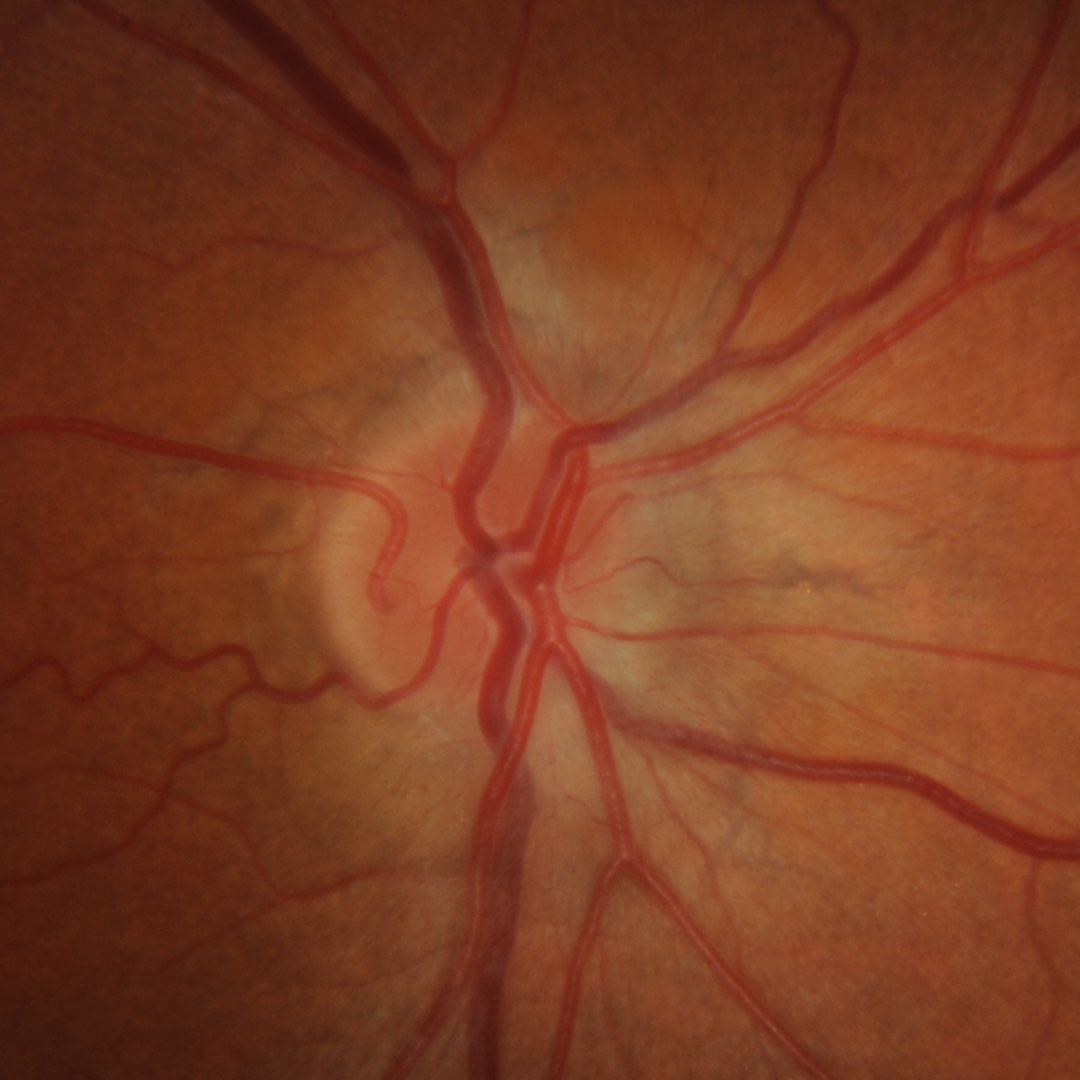
A 32-year-old Caucasian male with joint hypermobility and a family history of sarcoidosis. His best corrected visual acuity is 6/6 (20/20) in each eye.
-
Case 2
A 46-year-old Caucasian female with no known systemic conditions. Her best corrected visual acuity is 6/12 (20/40) in the right eye and 6/6 (20/20) in the left.
DIfferential Diagnoisis
References
Barteselli, G. Viola, F. (2015) Comet Lesions in Pseudoxanthoma Elasticum, Retina: May Volume 35 - Issue 5 - p 1051-1053.
Corbelli, E. Carnevali, A. Marchese, A. Cicinelli, M. Querques, L. Sacconi, R. Bandello, F. Querques, G. (2018) Optical coherence tomography angiography features of angioid streaks. Retina: November Volume 38 - Issue 11 - p 2128-2136.
Giachetti Filho, RG., Zacharias, LC., Monteiro, TV. et al. (2016) Prevalence of outer retinal tubulation in eyes with choroidal neovascularization. Int J Retin Vitr 2, 6 (2016).
Marchese, A., Parravano, M., Rabiolo, A. et al.(2017) Optical coherence tomography analysis of evolution of Bruch’s membrane features in angioid streaks. Eye 31, 1600–1605.