
Overview
Commotio retinae, the whitening and opacification of retina, occurs due to injury to the photoreceptor outer segments from blunt trauma to the eye. Vision loss and recovery varies depending on the severity of trauma, with milder cases resolving within a short period with no permanent effect on vision, while severe cases can cause permanent vision loss.
On OCT imaging, the region of commotio retinae corresponds to hyperreflectivity of the ellipsoid zone. Milder cases are transient and recover with disappearance of the retinal opacity over several days. However, in more severe cases of commotio retinae, there may be more disorganisation to the ellipsoid zone, hyperpigmentation, residual RPE changes and atrophy.
Case Examples
-
Case 1: Acute commotio retinae
A 23 year old male with a recent history of blunt trauma to the left eye. His best corrected visual acuity in this eye is 6/12+ (20/40+).
-
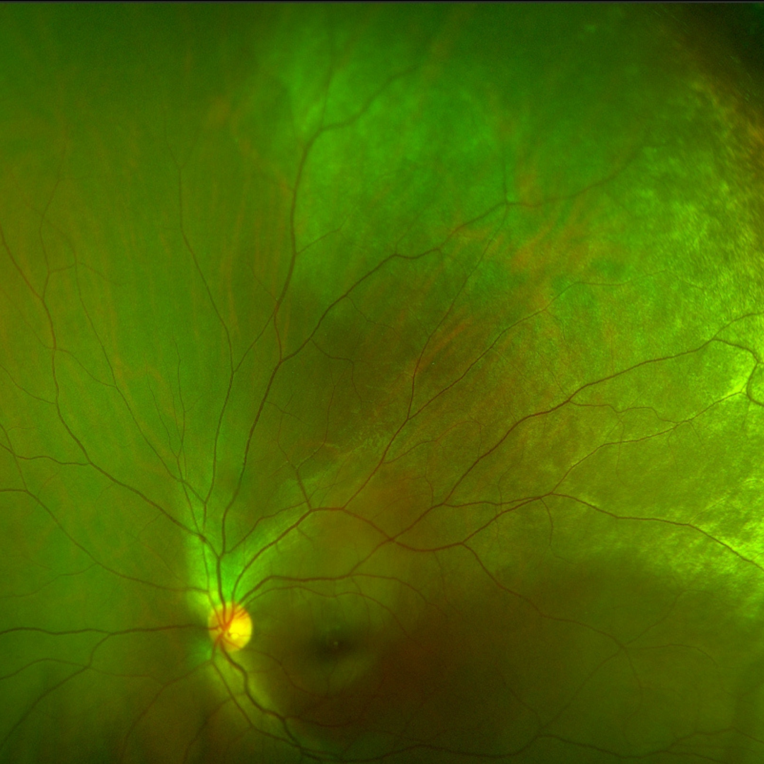
Case 2: Previous commotio retinae
A 27 year old Caucasian male with a history of blunt trauma to the right eye at the age of 6. At the time he recalls losing the vision in this eye but is unsure how long it took to return. His best corrected visual acuity in the right eye is 6/9.5- (20/32-).
Differential Diagnosis (acute)
Differential Diagnosis (Chronic)
References
Ahn, SJ, Woo, SJ. Kim, KE. Jo, DH, Ahn, J, Park, KH (2013) Optical Coherence Tomography Morphologic Grading of Macular Commotio Retinae and its Association With Anatomic and Visual Outcomes, American Journal of Ophthalmology, Vol 156(50) pp 994-1001.
Souza-Santos, F. Lavinsky, D. Moraes, N. Castro, A. Cardillo, J. Farah, M. (2012). Spectral-Domain Optical Coherence Tomography in Patients with Commotio Retinae. Retina, 32, pp 711-718.