
Overview
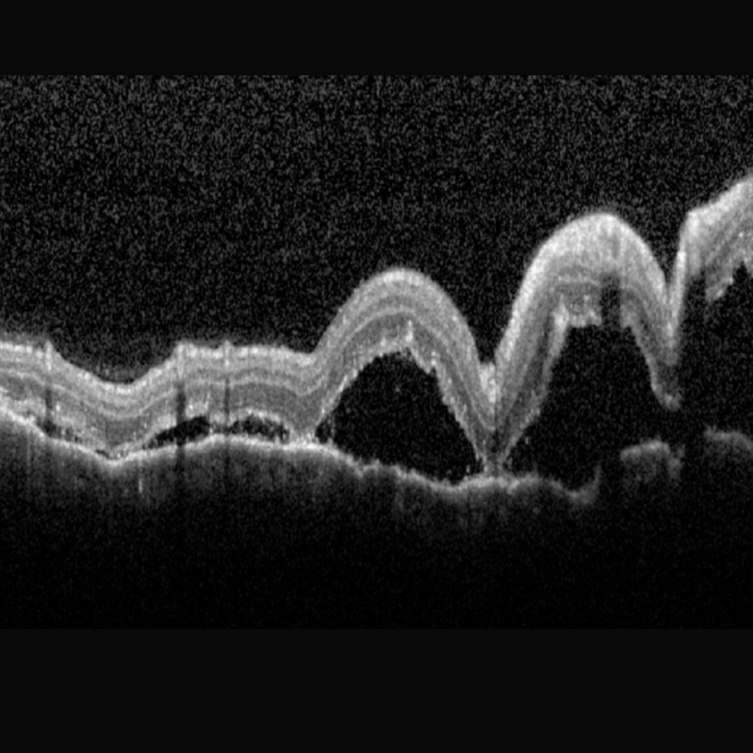
Uveal effusion occurs when fluid escapes from the choriocapillaris, causing choroidal engorgement. Fluid accumulates in the suprachoroidal space leading to choroidal thickening and detachment. Widespread exudative (serous) retinal detachment is typically associated. Often retinal exudates may be seen prior to the detachment occurring.
Uveal effusion may occur in association with ocular hypotony (surgical or traumatic cause) or inflammation (posterior scleritis or pars planitis).
Uveal effusion syndrome is a condition characterised by recurrent episodes of uveal effusion and is a diagnosis of exclusion. Chronic disease gives rise to RPE changes, giving the retina what is described as a “leopard spot” appearance. This syndrome is associated with hyperopia or nanophthalmia, but can also be idiopathic.
Case Example
-
Case 1
A 51 year old male who presents with red, sore eyes bilaterally for the last 3 days. Pinhole acuities were 6/30 (20/100) in the right eye and 6/15 (20/50) in the left. Pupil reactions were sluggish bilaterally with the right eye more affected than the left. There were cells in the anterior chamber.
Given the clinical presentation, this patient was referred urgently to an ophthalmologist where he was diagnosed with underlying posterior scleritis and treated appropriately. The scleritis was thought to be related to an underlying systemic connective tissue disorder.
Differential Diagnosis
References
Elagouz, M. Stanescu-Segall, D. Jackson, TL (2010) Uveal Effusion Syndrome. Survey of Ophthalmology, Volume 55, Issue 2, pp 134-145.
Shah, P. Yohendran, J. Hunyor, A. (2016). Uveal Effusion: Clinical Features, Management, and Visual Outcomes in a Retrospective Case Series. Journal of Glaucoma, 25, e329-e335