
Overview
The spectrum of white dot syndromes are characterised by hypopigmented inflammatory lesions of the outer retina, RPE, choriocapillaris and/or choroid.
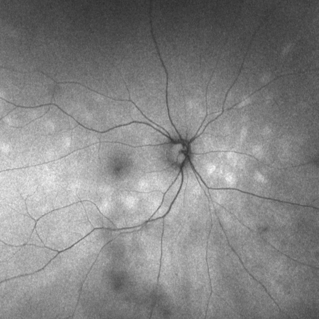
White dot syndromes have associated symptoms of blurred vision, floaters, photopsias, and scotoma and some also show retinal and/or vitreal signs of inflammation. Multimodal imaging is useful in distinguishing between the disease identities within the group of white dot syndromes.
Within this resource, we will explore the key white dot syndromes and illustrate the clinical characteristics of each using multimodal imaging.
Sub-Topics
Multiple evanescent white dot syndrome (MEWDS)
Learn more
Acute posterior multifocal placoid pigment epitheliopathy (AMPPE)
Learn more
Multifocal Choroiditis (MFC) and Punctate Inner Choroidopathy (PIC)
Learn more
Birdshot chorioretinopathy
Learn more
Serpiginous choroiditis
Learn more
Acute macular neuroretinopathy
Learn more
Acute zonal occult outer retinopathy (AZOOR)
Learn more