
Overview
Acute posterior multifocal placoid pigment epitheliopathy (APMPPE) is characterised by the presence of plaque-like lesions at the posterior pole which resolve over 1-2 weeks.
This condition affects men and women equally and typically starts off unilaterally with the second eye becoming involved a few days later. This condition resolves spontaneously without treatment and has a good prognosis unless there is recurrent disease, the fovea is affected or the patient is over 60 years of age. Large lesions may result in RPE atrophy and hyperplasia.
APMPPE can be associated with anomalous findings such as optic disc inflammation, periphlebitis, disc neovascularisation and exudative retinal detachment.
Presenting symptoms include blur, photophobia and paracentral scotoma. Clinically, cream-coloured placoid lesions are noted in the acute phase. OCT shows hyper-reflectivity of the outer retinal layer, disruption to the ellipsoid zone and/or RPE and sometimes outer retinal cysts.
As the lesion resolves, the hyper-reflectivity on OCT reduces and RPE atrophy may be noted.
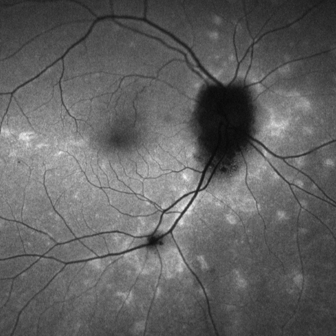
Fundus autofluorescence imaging shows the placoid lesions to be hypo-fluorescent in the acute phase, becoming hyper-fluorescent as they regress.
Case Examples
-
Case 1
An asymptomatic 59 year old Caucasian female with unaided visual acuities of 6/6- (20/20-) in each eye.
This case was also discussed in a paper by Wong et al (2015) Optom Vis Sci.
Fundus photographs (right and left eye)
More infoStereoscopic image (right optic disc)
More infoOptomap widefield (top) and green separation (bottom) images
More infoFundus autofluorescence images (right and left eye)
More infoSpectralis OCT line scan through the hypo-fluorescent lesion
More infoSpectralis OCT line scan through a hyper-fluorescent lesion
More infoSpectralis OCT volume and line scan (right optic nerve)
More infoB-scan ultrasound (right eye)
More info24-2 SITA Standard visual field
More info
Differential Diagnosis
References
Knickelbein, JE. Nida Sen, H. (2016) Multimodal Imaging of the White Dot Syndromes and Related Diseases J Clin Exp Ophthalmol. 2016 June ; 7(3).
Wong, E. Nivison-Smith, L. Assaad, NN. Kalloniatis, M. (2015). OCT and fundus autofluorescence enhances visualization of white dot syndromes. Optometry and Vision Science, 92(5):642-53.