
Overview
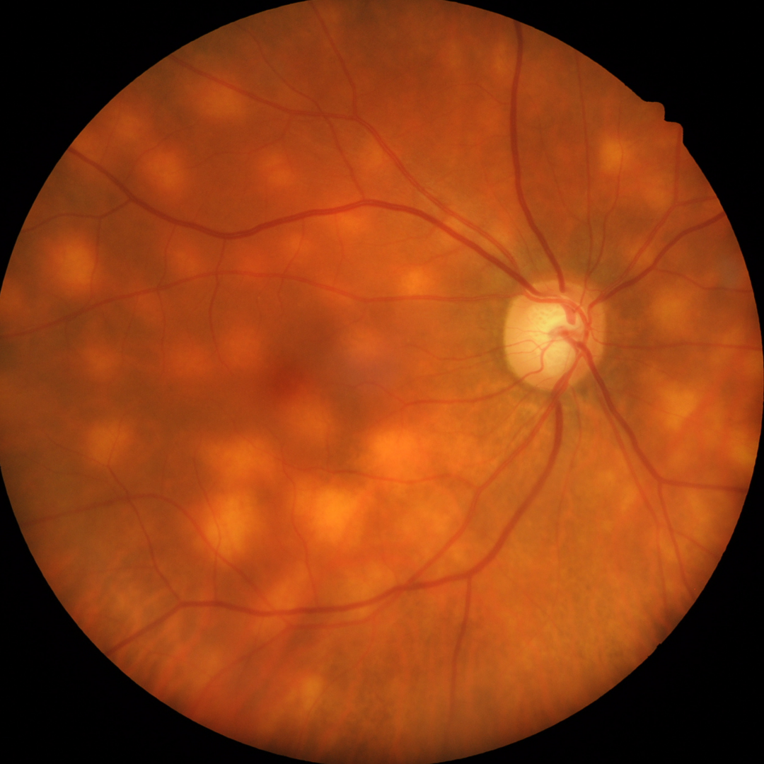
Birdshot chorioretinopathy is a bilateral posterior uveitic condition with characteristic hypo-pigmented round or oval lesions with indistinct borders that radiate out from the optic nerve. It primarily affects females aged between 40 and 70 years.
Presenting symptoms typically include blurred vision, nyctalopia, paracentral scotoma, photopsia and floaters.
Vitreous inflammation and retinal vasculitis are often present in this condition. The characteristic retinal lesions however usually appear later in the disease process so OCT imaging is important for early diagnosis.
OCT shows a loss of the Ellipsoid zone, outer retinal hyper-reflective loci, retinal thinning and RPE atrophy. Cystoid macular oedema and epiretinal membrane are also often present with macular oedema often causing reduced vision.
The choroid shows a thinning or loss of Sattler’s layer, focal hypo-pigmentation and may show a suprachoroidal hypo-reflective space.
Fundus autofluorescence (FAF) imaging shows hypo-fluorescent spots throughout the retina, however these areas are not aligned with the hypo-pigmented aeras seen clinically. There may be more hypo-flourescent areas than there are hypo-pigmented areas of retina, and not all the hypo-pigmented retinal areas produce hypo-fluorescent spots.
This condition is typically both chronic and recurrent.
Case Examples
-
Case 1
A 55 year old Caucasian female with best corrected visual acuity of 6/6- (20/20-) in each eye. She reports long-standing floaters but no other visual symptoms. Her referring optometrist notes that these lesions have been present and stable for at least 3 years.
Fundus photographs (right and left eye)
More infoOptomap widefield (1), red separation (2) and green separation (3) images - right eye
More infoOptomap widefield (1), red separation (2) and green separation (3) images - left eye
More infoFundus autofluorescence imaging (right and left eye)
More infoSpectralis OCT line scan (right macula)
More infoSpectralis OCT line scan (left macula)
More infoSpectralis OCT line scan (left superior macula)
More info30-2 SITA-standard visual field
More info
Differential Diagnosis
References
Knickelbein, JE. Nida Sen, H. (2016) Multimodal Imaging of the White Dot Syndromes and Related Diseases J Clin Exp Ophthalmol. 2016 June ; 7(3): .
Wong, E. Nivison-Smith, L. Assaad, NN. Kalloniatis, M. (2015). OCT and fundus autofluorescence enhances visualization of white dot syndromes. Optometry and Vision Science, 92(5):642-53.