
Overview
Multifocal choroiditis (MFC) and punctate inner choroidopathy (PIC) form a spectrum of conditions characterised by the presence of multiple small, circular inflammatory lesions within the RPE and choroid. Over time these yellow-white lesions develop a pigmented border.
This condition is most commonly bilateral and affects young females most frequently. Vision may be affected by epiretinal membrane, macular oedema, vitritis, choroidal neovascularisation or fibrotic scarring at the macula.
Acute MFC is typically accompanied by vitritis and often also inflammation in the anterior chamber. PIC is thought to be part of the spectrum of MFC and is a term used to describe a presentation limited to the posterior pole, consisting of small lesions and no vitritis or anterior chamber inflammation. Presumed ocular histoplasmosis syndrome is also considered to be part of this MFC spectrum.
OCT shows hyper-reflective material between the RPE and Bruch’s membrane with hyper-transmission of the signal through to the choroid. Sub-RPE deposits and disruption of the outer retinal layers may also be noted. Lesions appear hypo-fluorescent on FAF imaging, typically showing a border of hyper-fluorescence in the acute stages, but becoming completely hypo-fluorescent over time.
Case Examples
-
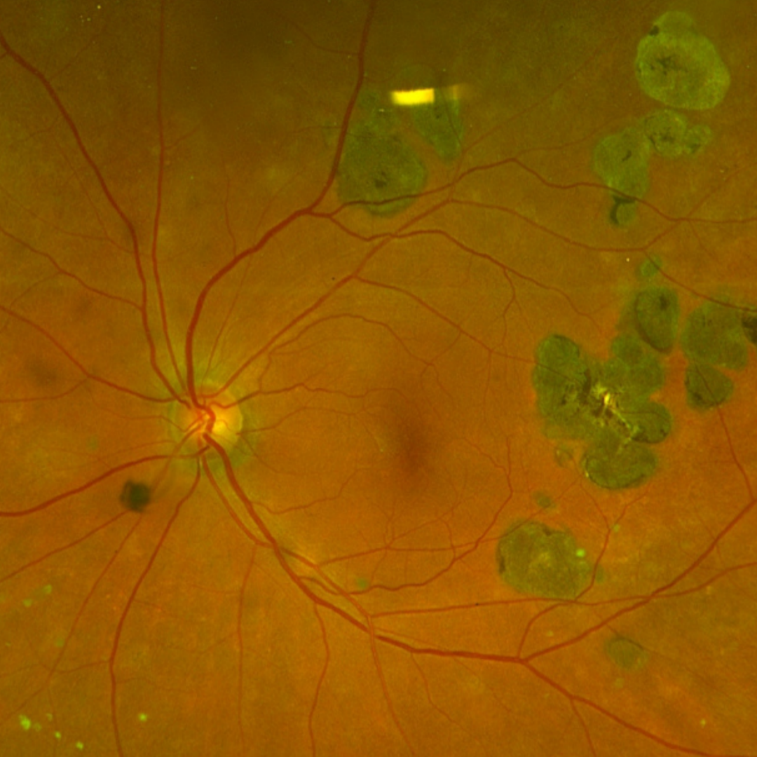
Case 1: Punctate inner choroidopathy
An asymptomatic 75 year old Caucasian male who has had an amblyopic right eye since he was young. His best corrected visual acuity is hand movements in the right eye and 6/6 (20/20) in the left.
Fundus photographs (right and left eye)
More infoRed-free images (right and left eye)
More infoOptomap widefield (1), red-separation (2) and green separation (3) images - right eye
More infoOptomap widefield (1), red-separation (2) and green separation (3) images - left eye
More infoFundus autofluorescence images (right and left eye)
More infoSpectralis OCT volume and line scans (right eye)
More info -
Case 2: Multifocal choroiditis
A 78 year old asymptomatic Asian female with best corrected visual acuity of 6/9 (20/30) in each eye.
Fundus photographs (right and left eye)
More infoRed-free images (right and left eye)
More infoOptomap widefield images (right and left eye)
More infoFundus autofluorescence images (right and left eye)
More infoCirrus OCT line scan (right macula)
More infoCirrus OCT line scans (left eye)
More info
DIfferential Diagnosis
References
Knickelbein, JE. Nida Sen, H. (2016) Multimodal Imaging of the White Dot Syndromes and Related Diseases J Clin Exp Ophthalmol. 2016 June ; 7(3): .
Wong, E. Nivison-Smith, L. Assaad, NN. Kalloniatis, M. (2015). OCT and fundus autofluorescence enhances visualization of white dot syndromes. Optometry and Vision Science, 92(5):642-53.