
Overview
Multiple evanescent white dot syndrome (MEWDS) is a unilateral, spontaneously resolving condition primarily affecting females between the ages of 20 and 45. It is often preceded by a virus.
Presenting symptoms include a sudden reduction in visual acuity, photopsia, visual scotomas (temporal or paracentral) and dyschromatopsia.
Clinically, multiple white dots may be seen throughout the retina and the fovea typically has a granular appearance. A mild vitritis and hypeaemic disc with or without blurred disc margins may also be noted in some cases.
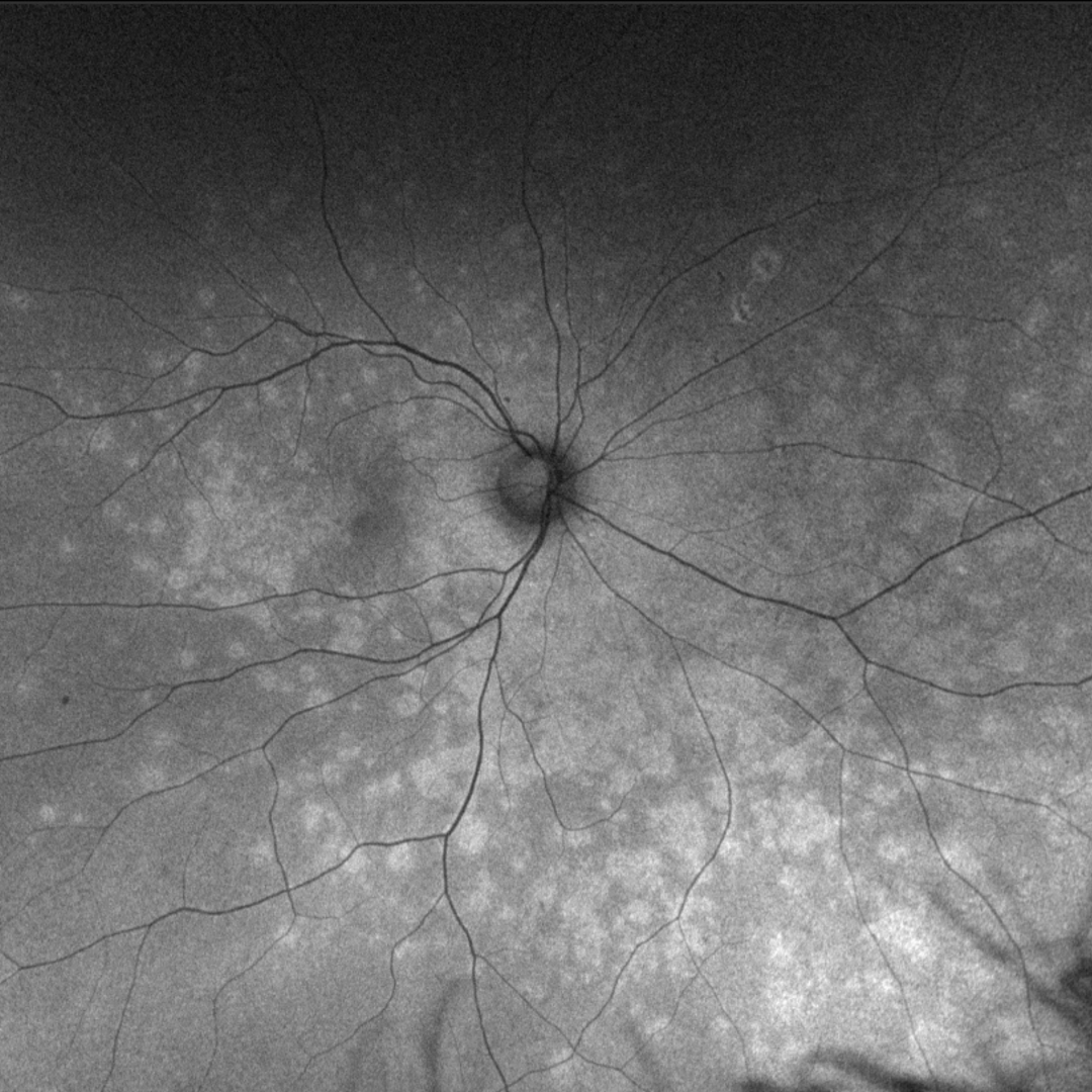
Fundus autofluorescence imaging shows hyper-fluorescence of the white retinal dots and pinpoint hypo-fluorescence in the area of foveal granularity.
OCT shows disruption of the ellipsoid zone in areas corresponding to the white dots, indicating that the photoreceptors are impacted in this disease.
MEWDS resolves spontaneously over a period of weeks to months with vision and visual fields returning to normal. This condition however may be recurrent, in which case atrophy of the photoreceptors may be noted.
Case Examples
-
Case 1: Acute MEWDS with resolution over 12 months
A 44 year old Caucasian female with best corrected visual acuity of 6/7.5 (20/25) in the right eye. She notes "spots" in her vision that do not move as well as blur when looking at text. These symptoms have been present for 3 weeks and at the time they appeared, she was unwell.
Optomap widefield (1), red separation (2) and green separation (3) images - initial visit
More infoFundus autofluorescence image (initial visit - right eye)
More infoCirrus OCT line scan (right superior retina)
More info24-2 SITA-standard visual field
More infoOptomap widefield (1), red separation (2) and green separation (3) images
More infoFundus autofluorescence image (follow-up visit - right eye)
More info -
Case 2: Resolving MEWDS
A 33 year old female with best corrected visual acuity of 6/6 (20/20) in the left eye. She has a recent history of a flu-like virus that lasted 10 months. She does not report any current visual symptoms
Differential Diagnosis
References
Knickelbein, JE. Nida Sen, H. (2016) Multimodal Imaging of the White Dot Syndromes and Related Diseases J Clin Exp Ophthalmol. 2016 June ; 7(3): .
Wong, E. Nivison-Smith, L. Assaad, NN. Kalloniatis, M. (2015). OCT and fundus autofluorescence enhances visualization of white dot syndromes. Optometry and Vision Science, 92(5):642-53.