
Overview
Gyrate atrophy is an autosomal recessive condition affecting the outer retina, RPE and choriocapillaris. It is caused by an accumulation of plasma ornithine in the retina which is toxic to the RPE and choroid.
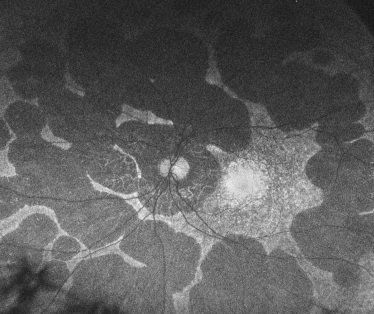
Early in the disease process, patients develop large peripheral areas of chorioretinal atrophy. These areas eventually coalesce forming a scalloped border peripherally, at the junction of the normal and abnormal retina.
Nyctalopia usually occurs in the first decade of life, followed by a progressive loss of visual field and eventually central vision is also lost.
The condition is associated with high myopia and astigmatism as well as early cataract formation. Related clinical findings may include macular oedema, choroidal neovascularisation and macular hole formation.
For most, there is no effective treatment.
Case Examples
-
Case 1
A 30-year-old Caucasian male with best-corrected visual acuity is 6/6 (20/20) in each eye.
Fundus photographs (right and left eye)
More infoOptomap widefield image (right and left eye)
More infoFundus autofluorescence images (right and left eye)
More infoSpectralis OCT volume (1) and line (2) scans - right eye
More infoSpectralis OCT volume (1) and line (2) scans - left eye
More info24-2 SITA Standard visual field
More info
Differential diagnosis
References
Sen,S. Kannan, SK., Shanmugam, U., Rajan, R., Babu, N., Vanniarajan,A. (2021) Variable phenotypes of gyrate atrophy in siblings with a nonsense mutation in OAT gene. Ophthalmic Genetics 0:0, pages 1-4.
Zhioua Braham, I., Ammous, I., Maalej, R. et al. (2018) Multimodal imaging of foveoschisis and macular pseudohole associated with gyrate atrophy: a family report. BMC Ophthalmol 18, 89