
Overview
White without pressure (WWOP) appears as an opaque area of white/grey retina, sometimes with a red-brown border. The appearance and distribution of these areas can change over time.
Studies have shown that the reflectivity changes seen in WWOP are not due to photopigment variations (Fawzi et al. 2014). While OCT imaging studies shows that WWOP corresponds to hyper-reflectivity at the level of the outer retina, specifically the outer segment and ellipsoid zones, the potential contribution of vitreoretinal traction has not yet been excluded.
Dark without pressure (DWOP) is most commonly found in darkly pigmented retinas. It presents as a flat, dark patch of the retina with well defined, sometime scalloped borders. Similar to WWOP it can change over time. DWOP has hypo-reflective changes in the same layers as WWOP.
Case Examples
-
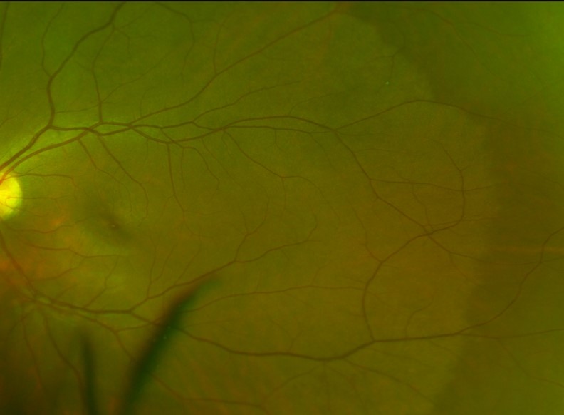
Case 1
A 29-year-old asymptomatic Asian male with best corrected visual acuity of 6/6 (20/20) in the left eye.
-
Case 2
A 28-year-old asymptomatic Asian female with best corrected visual acuity in each eye of 6/6 (20/20).
Optomap pseudocolour (1), green separation (2) and fundus autofluorescence (3) images - right eye
More infoOptomap (1), green separation (2) and fundus autofluorescence (3) images - left eye
More infoSpectralis OCT line scans through an area of dark without pressure (nasal periphery, left eye)
More info
Differential Diagnosis
References
Diaz, Rocio, Sigler, Eric, Randolph, John, Rafieetary, Mohammad & Calzada, Jorge. (2014). Spectral Domain Optical Coherence Tomography Characteristics of White-Without-Pressure. Retina, 34, 1020-1021.
Fawzi AA, Nielsen JS, Mateo-Montoya A, Somkijrungroj T, Li HK, Gonzales J, Mauget-Faÿsse M, Jampol LM. (2014) Multimodal imaging of white and dark without pressure fundus lesions. Retina. Dec;34(12):2376-87.