
Overview
Peripheral cystoid degeneration is characterised by clusters of tiny vesicles within a hazy grey area of thickened retina extending from the ora serrata. There may be small red cysts within areas of cystoid degeneration, which may mimic the appearance of atrophic retinal holes.
OCT imaging shows hyporeflective cysts within the sensory retina. Coalescence of cysts may occur and is thought to be a precursor to development of retinoschisis.
Case Examples
-
Case 1
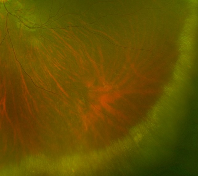
A 31-year-old asymptomatic Asian female with best corrected visual acuity of 6/6 (20/20) in each eye.
Optomap pseudocolour (1), green separation (2) and fundus autofluorescence (3) images - right eye, temporal retina
More infoOptomap pseudocolour (1), green separation (2) and fundus autofluorescence (3) images - left eye, temporal retina
More infoOptomap pseudocolour (1) and green separation (2) images - left eye, inferior retina
More infoSpectralis OCT line scan
More info -
Case 2
A 44-year-old asymptomatic Asian female with best corrected visual acuity of 6/6 (20/20) in each eye.
-
Case 3
A 43-year-old asymptomatic Asian female with best corrected visual acuity of 6/6 (20/20) in each eye.
-
Case 4
A 31-year-old asymptomatic Caucasian female with best corrected visual acuity of 6/6 (20/20) in each eye.
Differential Diagnosis
References
Hilel Lewis (2003) Peripheral retinal degenerations and the risk of retinal detachment. American Journal of Ophthalmology, Volume 136, Issue 1. Pages 155-160,
Thanos, A., Todorich, B., Pasadhika, S., Khundkar, T., Xu, D., Jain, A., Ung, C., Faia, L. J., Capone, A., Williams, G. A., Yonekawa, Y., Sarraf, D., & Wolfe, J. D. (2019). Degenerative Peripheral Retinoschisis: Observations From Ultra-Widefield Fundus Imaging. Ophthalmic Surgery, Lasers & Imaging Retina, 50(9), 557-564.