
Overview
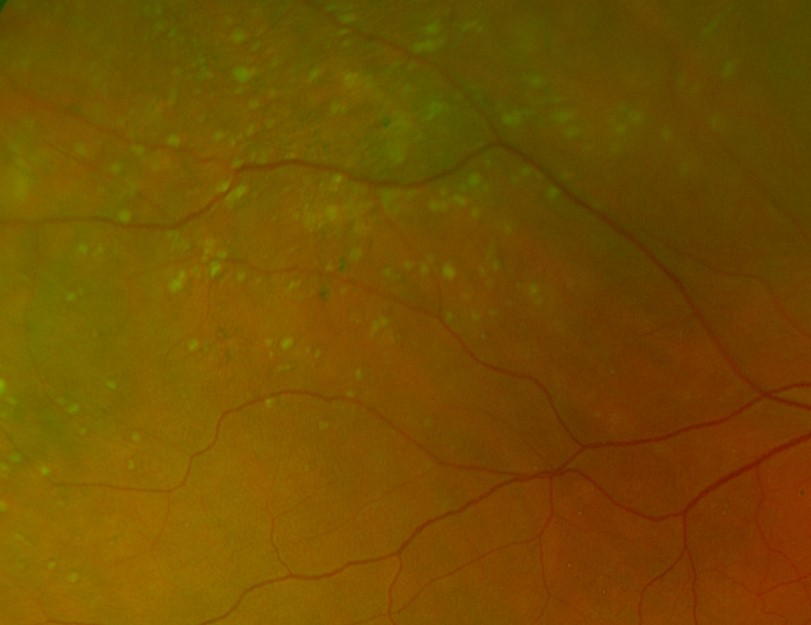
Peripheral drusen present as small, focal, yellow lesions in the retinal periphery, often near the equator. Drusen may have pigmented borders and are often associated with reticular pigmentary degeneration (more information on this condition is available on the link at the bottom of this page).
Peripheral drusen are usually bilateral.
OCT shows elevations of the RPE with homongenous reflectivity underneath the elevations.
Some authors have shown an association between peripheral drusen and the drusen of AMD, with the suggestion made that peripheral drusen may be a sign of genetic susceptibility to AMD, even in those who do not show signs of the macular condition. These conclusions are still somewhat contentious in the literature however.
Case Examples
Differential Diagnosis
References
Domalpally A, et al. (2017) Peripheral Retinal Changes Associated with Age-Related Macular Degeneration in the Age-Related Eye Disease Study 2: Age-Related Eye Disease Study 2 Report Number 12 by the Age-Related Eye Disease Study 2 Optos PEripheral RetinA (OPERA) Study Research Group. Ophthalmology. Apr;124(4):479-487.
Johanna M. Seddon, Robyn Reynolds, Bernard Rosner; Peripheral Retinal Drusen and Reticular Pigment: Association with CFHY402H and CFHrs1410996 Genotypes in Family and Twin Studies. (2009) Invest. Ophthalmol. Vis. Sci. 2009;50(2):586-591.
Nivison-Smith L, Milston R, Chiang J, Ly A, Assaad N, Kalloniatis M. (2018) Peripheral retinal findings in populations with macular disease are similar to healthy eyes. Ophthalmic Physiol Opt. Nov;38(6):584-595.