
Overview
Peripheral reticular pigmentary degeneration (also known as senile reticular pigmentary degeneration) is characterised by reticular or net-like pigmentary changes that form geometric patterns in the peripheral retina, often accompanied by peripheral drusen. These may appear similar to the bone-spicules in retinitis pigmentosa. However, unlike retinitis pigmentosa, it does not impact visual function or the visual field.
Fundus changes are typically bilateral and superonasal in location, however, it can extend to the temporal retina in some cases. The incidence of PRPD increases with age.
OCT scans through the areas of reticular change typically show thickening of the RPE.
Recent fluorescein angiography studies indicated that ischemia of the choriocapillaris secondary to vascular insufficiency is a possible cause of this degenerative change. Some studies have suggested an association between AMD and PRPD, although this inconclusive.
Case Examples
-
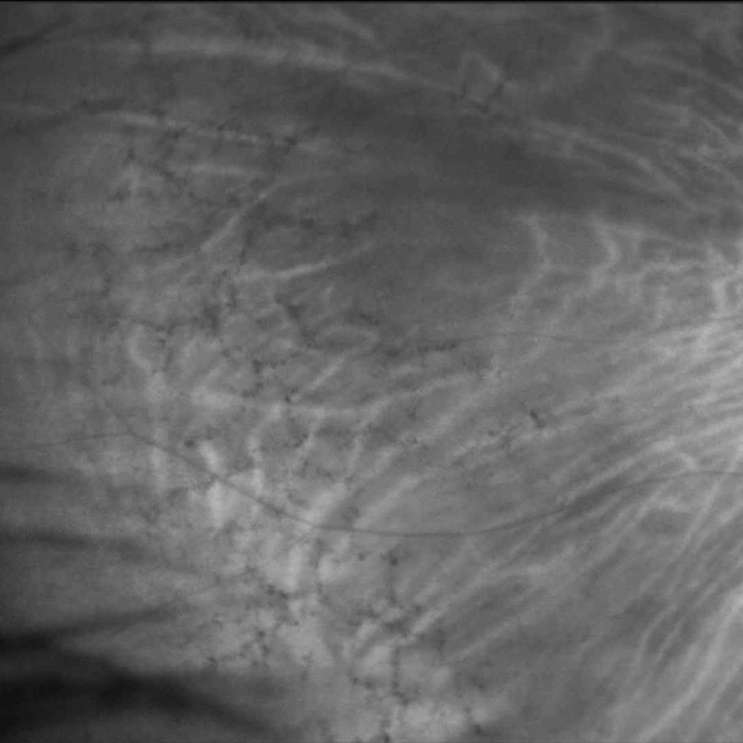
Case 1
A 72 year old Caucasian female with best corrected visual acuities of 6/7.5 (20/25) in each eye.
Differential diagnosis
Additional differentials to consider include pigmentary changes secondary to trauma or inflammation.
References
Bae, K., Cho, K., Kang, S. W., Kim, S. J., & Kim, J. M. (2017). Peripheral Reticular Pigmentary Degeneration and Choroidal Vascular Insufficiency, Studied by Ultra Wide-Field Fluorescein Angiography. PloS one, 12(1), e0170526