
Overview
Atrophic holes are caused by thinning of the retina over time and are not associated with vitreoretinal traction. Chronic retinal atrophy leads to a round or oval shaped break full thickness break in the neurosensory retina.
Atrophic holes can range in size from tiny pinpoint lesion through to as large as 2 disc diameters in size. They may be associated with surrounding subretinal fluid (a fluid cuff) which can also be described as a subclinical retinal detachment. As with other retinal breaks, the presence of pigmentation surrounding an atrophic hole is a sign of chronicity.
Holes may occur internal to areas of lattice degeneration or snailtrack degeneration as these lesions are characterised by retinal thinning. They may also occur independent of any pre-disposing lesion. Most atrophic holes are solitary however multiple holes can occur within areas of lattice or snailtrack degeneration.
Small atrophic holes can develop after blunt trauma to the eye, in areas of commotio retinae.
There are no other generally accepted risk factors for atrophic holes and it is estimated that the prevalence of this finding is approximately 5%.
Case Examples
-
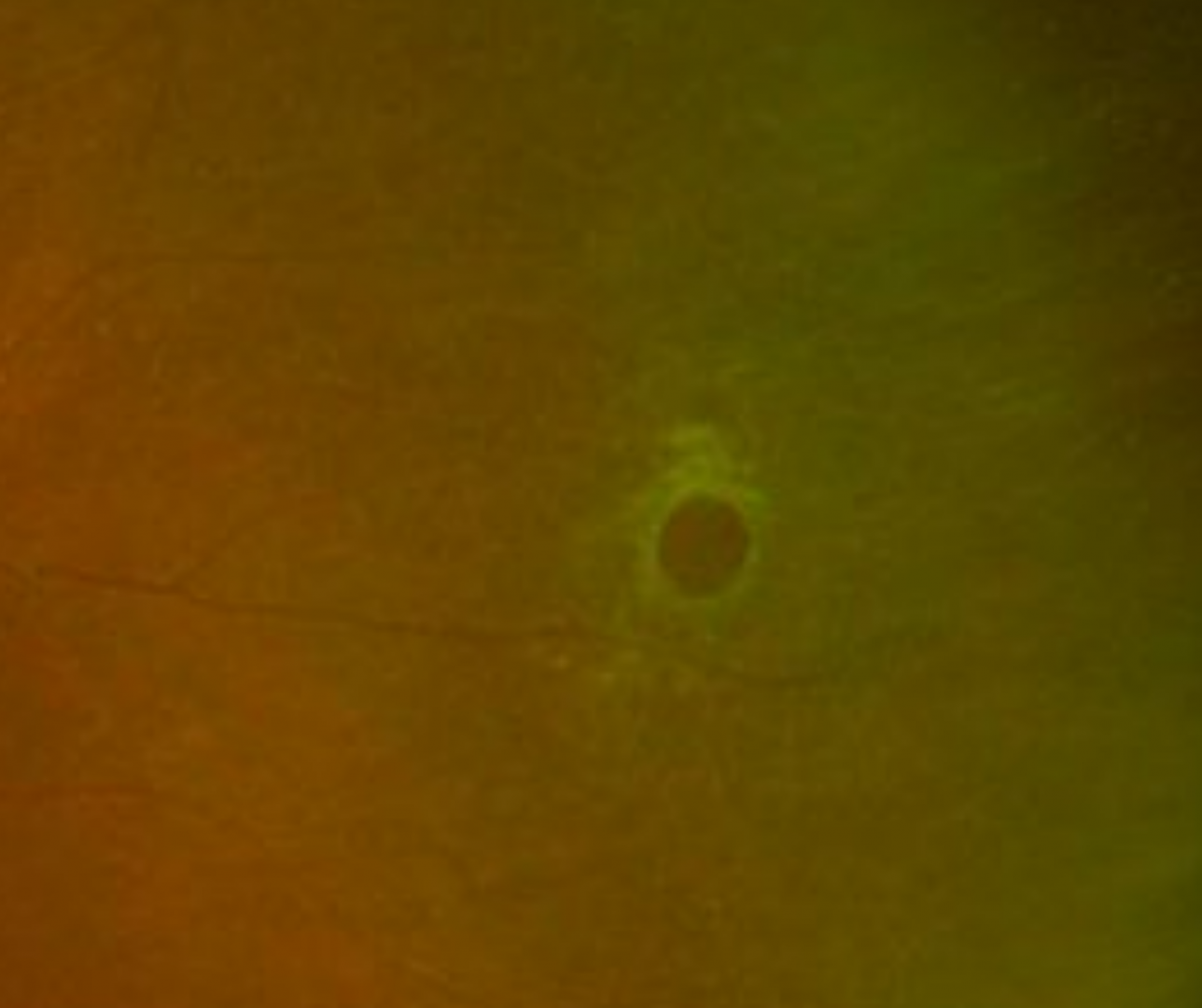
Case 1
An asymptomatic 34 year old Caucasian female with high myopia and best corrected visual acuity of 6/6 (20/20) in the left eye.
-
Case 2
A 29 year old asymptomatic Indian female with high myopia and best corrected visual acuity of 6/6 (20/20) in the left eye.
-
Case 3
A 75 year old Asian male with best corrected visual acuity in the left eye of 6/9 (20/30).
Differential diagnosis
Associated Topics
References
Atrophic Retinal Holes. In: Schmidt-Erfurth U., Kohnen T. (eds) (2018) Encyclopedia of Ophthalmology. Springer, Berlin, Heidelberg.
H. Dunbar Hoskins Jr., MD Center for Quality Eye Care (2019) Posterior Vitreous Detachment, Retinal Breaks, and Lattice Degeneration Preferred Practice Pattern. American Academy of Ophthalmology.
Wilkinson,C. and Cochrane Eyes and Vision Group Cochrane Database Syst Rev. (2014) Sep; 2014(9): CD003170