
Overview
As the eyes moves, the mobile vitreous exerts tractional forces on the retina. Over time, the vitreous liquifies and this internal movement becomes greater. Eventually, the vitreous undergoes a process of separation from the retina called posterior vitreous detachment (PVD). A retinal break may occur in areas of strong vitreoretinal attachment, in some cases creating a horseshoe tear.
In a horseshoe tear, the retinal flap remains attached to the vitreous and as the PVD continues to progress, the flap may be held open by this traction. This enables the movement of vitreous fluid into the subretinal space, causing separation of the RPE and neurosensory retina. Saccadic eye and head movements further lift the flap, leading to progression of the rhegmatogenous retinal detachment (RRD).
At least 50% of symptomatic horseshoe tears lead to RRD unless treated. Approximately 5% of asymptomatic horseshoe tears progress to retinal detachment.
Horseshoe tears are associated with areas of strong, persistent vitreoretinal attachment, including areas of lattice degeneration and vitreoretinal tufts. For more information about the pre-disposing lesions, please click on the links at the bottom of this page.
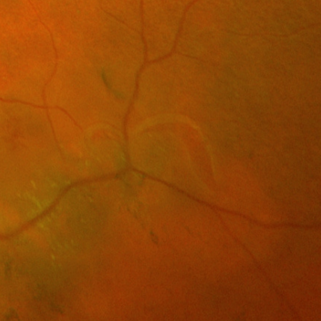
Clinically, a horseshoe tear presents as a red u-shaped or linear lesion surrounded by white-grey retinal tissue (fluid cuff) and in some cases associated with retinal detachment. When horseshoe-shaped, the apex of the tear points towards the posterior pole.
OCT imaging shows a retinal flap that is still attached to the vitreous with adjacent full thickness retinal break.
Urgent referral of an acute horseshoe tear is required.
Case Examples
-
Case 1
A 56-year-old Asian female who has noticed increased floaters in her right eye for the last 1-2 weeks. She is a moderate myope (-3.50DS in both eyes) and has best corrected visual acuity of 6/6 (20/20) in each eye.
-
Case 2
A 68-year-old Asian male with recent onset flashes of light noted in his left eye. His best corrected visual acuity in this eye is 6/6 (20/20).
-
Case 3
An 80-year-old asymptomatic Caucasian male who had cataract surgery 10 years prior. His best corrected visual acuity is 6/7.5 (20/20) in the left eye.
References
Ghazi, N., Green, W. Pathology and pathogenesis of retinal detachment. Eye 16, 411–421 (2002).
Wilkinson,C. and Cochrane Eyes and Vision Group Cochrane Database Syst Rev. (2014) Sep; 2014(9): CD003170.