
Overview
As the eyes moves, the mobile vitreous exerts tractional forces on the retina. Over time, the vitreous liquifies and this internal movement becomes greater. Eventually, the vitreous undergoes a process of separation from the retina called posterior vitreous detachment (PVD). In areas of strong vitreoretinal attachment, avulsion of the retina may occur, alleviating vitreoretinal traction and resulting in an operculated retinal hole. The fact that there is no ongoing traction means that an operculated hole is less likely to progress to RRD than a horseshoe retinal tear which has ongoing vitreoretinal traction.
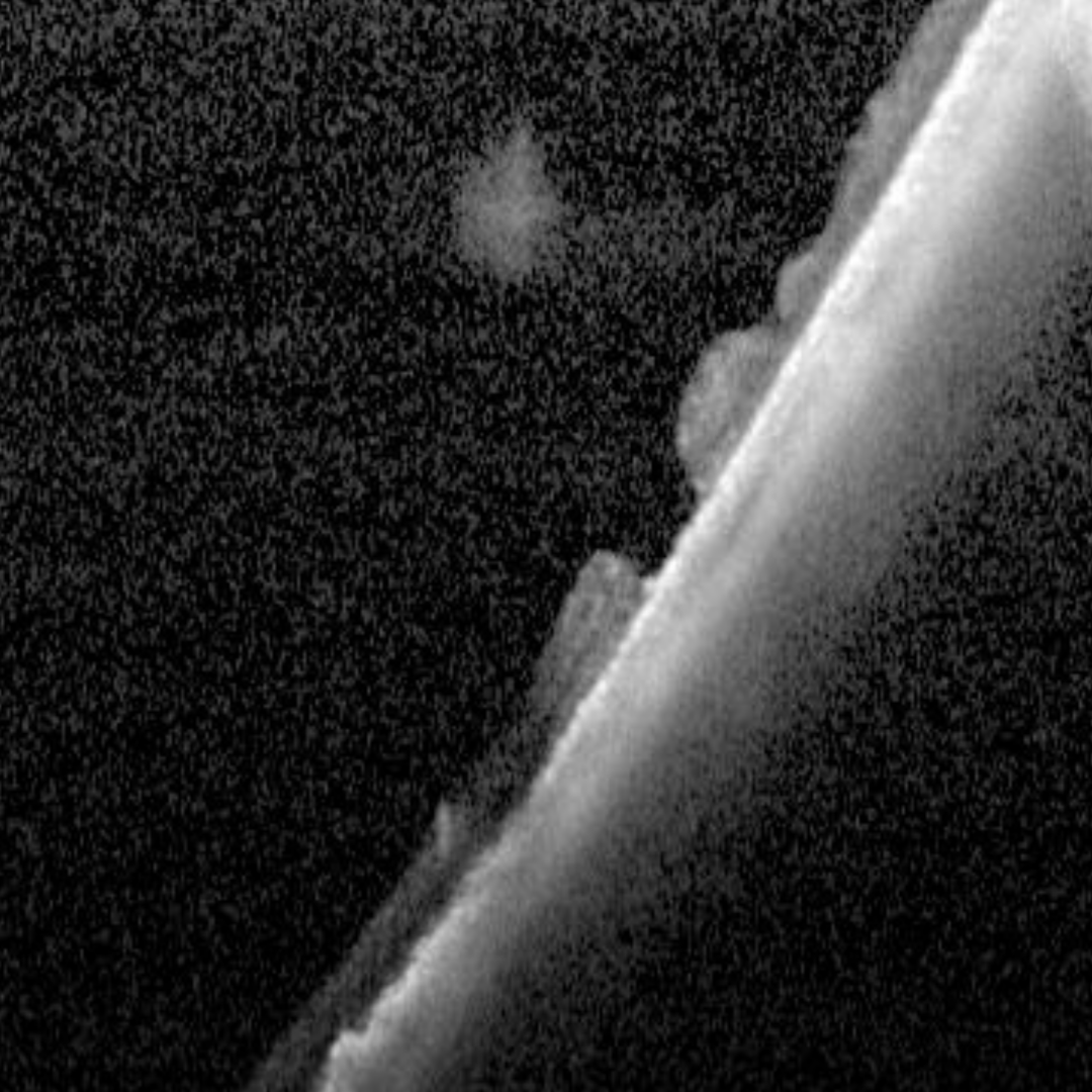
Operculated retinal holes appear as a round or oval-shaped red area in the retina with an overlying floating fragment of tissue (an operculum). When newly formed, the fragment is the same size as the hole underneath however over time it atrophies and will appear smaller than the section of missing retina. It may also be displaced away from the location of the hole with time.
Operculated retinal holes may be associated with subclinical retinal detachment (a fluid cuff). As with other retinal breaks, RPE hyperplasia is a sign of chronicity.
OCT shows a full thickness retinal break with the operculum often visible in the vitreous overlying the hole. The retinal hole may have subretinal fluid adjacent.
Operculated retinal holes rarely lead to a clinical retinal detachment.
Case Examples
-
Case 1: Operculated hole
An 83-year-old asymptomatic Caucasian male with best corrected visual acuity of 6/7.5 (20/25) in the left eye.
-
Case 2: Partial thickness operculated hole
A 31-year-old Middle Eastern female with moderate myopia and best corrected visual acuity of 6/6 (20/20) in the right eye.
-
Case 3: Operculated hole
An asymptomatic 66-year-old Asian male with best corrected visual acuity in the left eye of 6/6 (20/20).
Differential Diagnosis
References
H. Dunbar Hoskins Jr., MD Center for Quality Eye Care (2019) Posterior Vitreous Detachment, Retinal Breaks, and Lattice Degeneration Preferred Practice Pattern. American Academy of Ophthalmology.
Fraser, S., & Steel, D. (2010). Retinal detachment. BMJ clinical evidence, 2010, 0710.
Ghazi, N., Green, W. (2002) Pathology and pathogenesis of retinal detachment. Eye 16, 411–421.