
Overview
Retinal dialysis is most commonly associated with ocular trauma in young people. It occurs when the retina is separated from the pars plana at the ora serrata, leading to a circumferential retinal break.
Retinal dialysis occurs within the vitreous base. It can be confused with a giant retinal tear which is also associated with trauma and retinal detachment, but a giant retinal tear occurs posterior to the vitreous base insertion.
Case Example
-
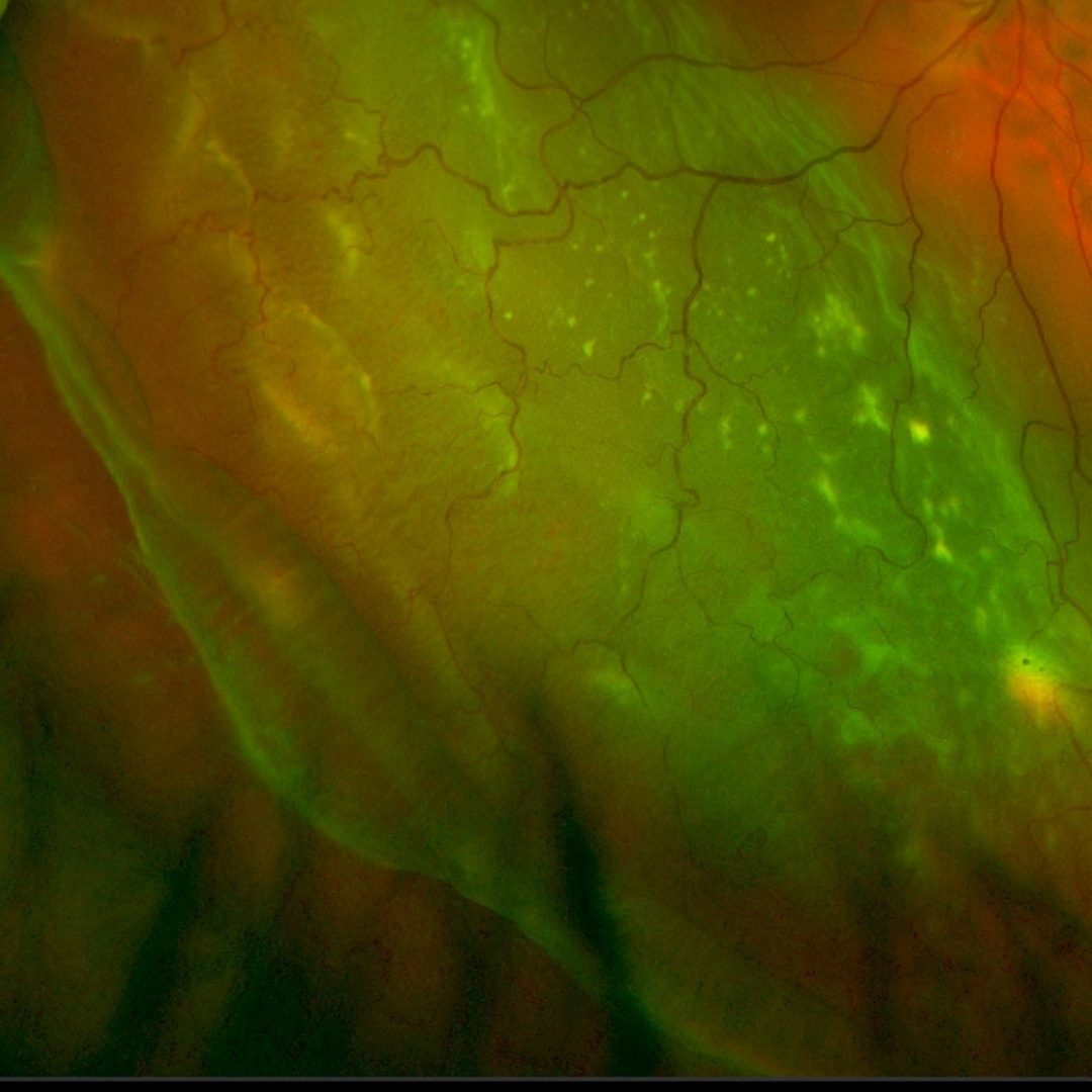
Case 1
A 22-year-old male with a history of blunt trauma to his right eye. He reports experiencing blurry vision since that incident approximately 2 years prior. His best corrected visual acuity is 6/38-1 (20/125-1) in the right eye.
Differential Diagnosis
References
Chang, J. S., Marra, K., Flynn, H. W., Jr, Berrocal, A. M., & Arroyo, J. G. (2016). Scleral Buckling in the Treatment of Retinal Detachment Due to Retinal Dialysis. Ophthalmic surgery, lasers & imaging retina, 47(4), 336–340.
H. Dunbar Hoskins Jr., MD Center for Quality Eye Care (2019) Posterior Vitreous Detachment, Retinal Breaks, and Lattice Degeneration Preferred Practice Pattern. American Academy of Ophthalmology.