
Overview
A typical retinoschisis presents as retinal splitting within the outer plexiform layer and is thought to occur due to the coalescence of cysts in peripheral cystoid degeneration. A typical retinoschisis is shallow, round or oval in shape and is found anterior to the equator. They are usually bilateral and are most commonly found in the inferotemporal retina.
Shallow retinoschisis may be difficult to detect, particularly on non-stereoscopic widefield imaging. Some clues include the presence of a mild haze partially obscuring choroidal detail and diversion of the blood vessels due to elevation of the inner retinal layers. Retinal blood vessels within an area of retinoschisis may also appear darker in colour than surrounding vessels.
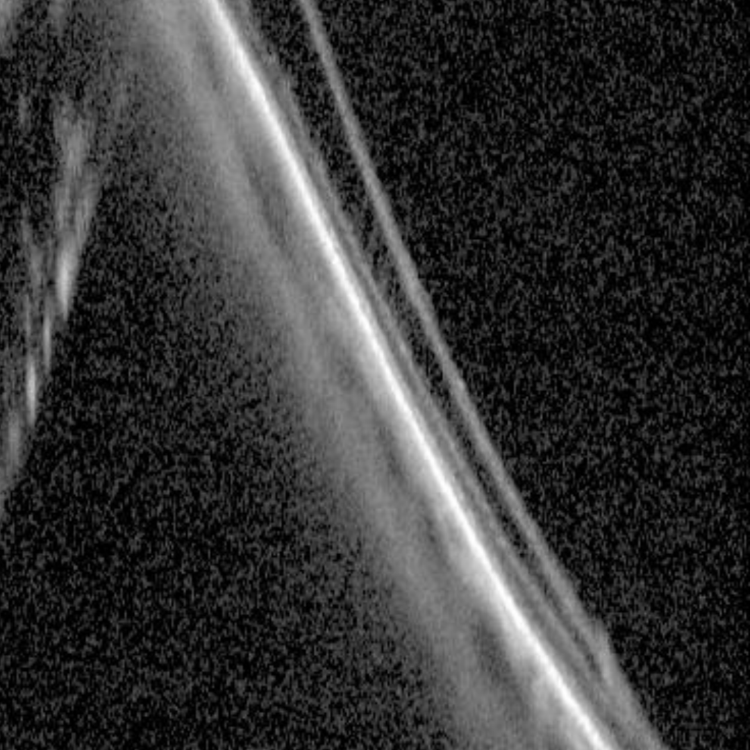
OCT shows retinal splitting at the level of the outer plexiform layer.
Other associated clinical findings may include areas of capillary non-perfusion, telangiectasia and retinal haemorrhages. Retinal holes or detachments are rare in the typical form of retinoschisis.
Case Examples
-
Case 1
A 47 year old asymptomatic Asian female with best corrected visual acuity of 6/6 (20/20) in the right eye.
Optomap and green separation images (right inferotemporal retina)
More infoSpectralis OCT line scans taken through (1) and at the border (2) of the area of cystoid degeneration
More infoOptomap and green separation images (left inferotemporal retina)
More infoSpectralis OCT line scans taken through (1) and at the border (2) of the area of cystoid degeneration
More info -
Case 2
An asymptomatic 70 year old Caucasian female with best corrected visual acuity of 6/6 (20/20).
References
Thanos, A., Todorich, B., Pasadhika, S., Khundkar, T., Xu, D., Jain, A., Ung, C., Faia, L. J., Capone, A., Williams, G. A., Yonekawa, Y., Sarraf, D., & Wolfe, J. D. (2019). Degenerative Peripheral Retinoschisis: Observations From Ultra-Widefield Fundus Imaging. Ophthalmic Surgery, Lasers & Imaging Retina, 50(9), 557-564.
Wallsh, JO, Gallemore,RP (2017) Identifying rhegmatogenous detachments in bullous retinoschisis with optical coherence tomography studies, American Journal of Ophthalmology Case Reports, Volume 6, Pages 38-40