
Overview
An astrocytic hamartoma is a benign tumour of the retinal nerve fibre layer and is composed of glial cells, namely astrocytes. They can arise from the retina, macula or optic nerve. Astrocytic hamartomas can present as an isolated finding but it has been shown to have systemic associations such as tuberous sclerosis and neurofibromatosis. They do not typically progress however in rare circumstances they may show rapid growth leading to exudative retinal detachments and necrosis.
Funduscopically, they present as either a well defined yellow-white elevation or as a flat, semi-translucent lesion with soft margins. They can be solitary or multiple. Over time calcification can occur resulting in a multinodular, ‘mulberry’ appearance.
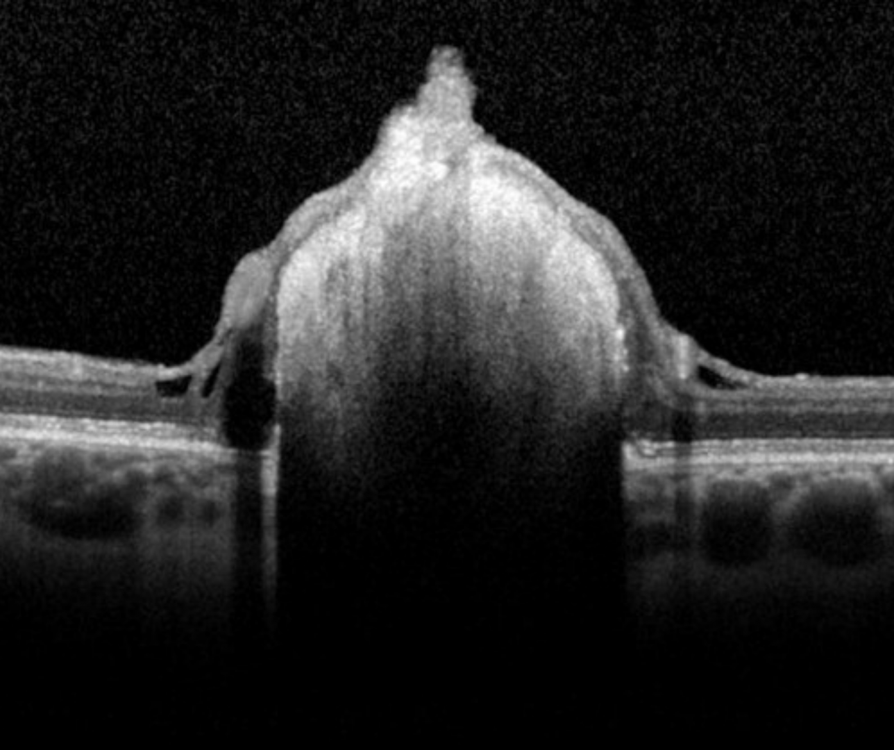
OCT imaging shows an intraretinal hyper-reflective elevation with posterior shadowing. With increasing calcification, moth eaten spaces can be seen internally.
Astrocytic hamartomas are typically hypo-autofluorescent on fundus autofluorescence imaging but can become hyper-autofluorescent as it becomes calcified.
In calcified types, the B-scan ultrasound will show a hyperacoustic signal with posterior shadowing.
Case Examples
-
Case 1: Retinal astrocytoma
A 51 year old asymptomatic Caucasian male with best corrected visual acuity of 6/4.8 (20/15) in the left eye.
-
Case 2: Retinal astrocytoma
An asymptomatic 48 year old Caucasian male with best corrected visual acuity of 6/6 (20/20) in the left eye.
-
Case 3: Juxtapapillary astrocytoma
A 24 year old Caucasian male with best corrected visual acuity of 6/4.8 in the left eye.
This patient reports no significant medical history, however given the possible association of astrocytoma with systemic conditions such as tuberous sclerosis, further medical investigations are warranted.
Differential diagnosis
References
Ciardella et al., 2009. Spectral Domain optical coherence tomography and autofluorescence findings in retinal astrocytic hamartomas.
Martin et al., 2009. Retinal astrocytic hamartoma