
- Overview (Bear Tracks)
- Overview (Polar Bear Tracks)
- Case Examples
- Differential Diagnosis (Bear Tracks)
- Differential Diagnosis (Polar Bear Tracks)
- References
Overview (Bear Tracks)
Congenital grouped pigmentation of the RPE (CGP-RPE) and its non-pigmented variation, grouped congenital albinotic retinal pigment epithelial spots (GCARPES) are congenital small, flat RPE lesions that may affect any part of the retina, but typically spares the macula.
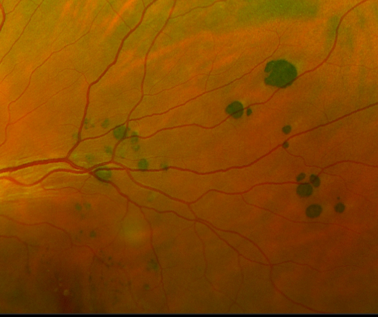
CGP-RPE are characterised by groups of small, well defined pigmented lesions that mimic animal footprints or paws. This condition is also termed as ‘bear tracks’. They can be found in a single sector or quadrant of the fundus and its size typically increases towards the periphery.
OCT imaging through the CGP-RPE can be variable. Some may show no detectable change but some may display RPE thickening, posterior shadowing with conformational elevation of the ellipsoid zone.
FAF typically shows a uniform hypo-autofluorescent signal. However, hyper autofluorescence has also been described which suggests RPE dysfunction or developing atrophy.
Overview (Polar Bear Tracks)
Related to CGP-RPE are grouped congenital albinotic retinal pigment epithelial spots (GCARPES).
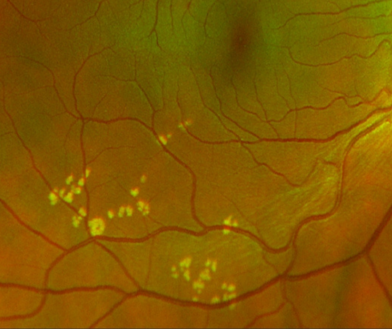
Funduscopic features of GCARPES include groups of small, flat hypopigmented lesions which can be distributed in a single sector or quadrant of the fundus. Its distribution is similar to CGP-RPE and is commonly referred to as polar bear tracks.
OCT imaging can show focal RPE thickening and disruption of the ellipsoid zone
FAF imaging can display both hyper and hypo autofluorescence.
Case Examples
-
Case 1: Bear tracks
An asymptomatic 32 year old Caucasian female with best corrected visual acuity of 6/6 (20/20) in the left eye.
This case is also discussed in the paper by Wang et al.(2020) Clin Exp Optom.
Fundus photograph (left eye)
More infoOptomap (1), red separation (2) and green separation (3) images - superior temporal retina, left eye
More infoFundus autofluorescence imaging (left eye)
More infoSpectralis OCT line scan through a lesion
More infoSpectralis OCT volume scan through several lesions
More info -
Case 2: Bear tracks
An asymptomatic 30 year old Caucasian male with best corrected visual acuity of 6/4.8 (20/15) in the left eye.
-
Case 3: Subtle bear tracks
An asymptomatic 29 year old Caucasian female with best corrected visual acuity of 6/4.8 (20/15) in the left eye
-
Case 4 : Polar bear tracks
An asymptomatic 29 year old Caucasian female with best corrected visual acuity of 6/4.8- (20/15-) in the right eye
Differential Diagnosis (Bear Tracks)
Differential Diagnosis (Polar Bear Tracks)
References
Ly, A. Nivison-Smith, L. Hennessy, M. Kalloniatis, M. (2015) Pigmented Lesions of the Retinal Pigment Epithelium, Optometry and Vision Science: Volume 92 - Issue 8.
Raval et al., 2019. Multimodal imaging of congenital hypertrophy of the retinal pigment epithelium (CHRPE) lesions at different presentations
Wang et al., 2020. Multimodal imaging characteristics of congenital grouped hyper- and hypo-pigmented fundus lesion. Clin Exp Optom, 103: 641-647.