
Overview
CHRPE is a benign, flat proliferation of retinal pigment eptithelial cells. It exists in 3 forms - typical, grouped and atypical CHRPE. This section will cover the typical type which presents as a solitary lesion. There is a link at the bottom of this page for further information about grouped and atypical CHRPE.
Typical CHRPE presents as a solitary, flat grey to black lesion with sharply demarcated margins on funduscopic examination. It is almost always unilateral and is located around the equator. They may have atrophied window-like defects called lacunae and can be surrounded by a halo of depigmentation.
OCT features include RPE thickening and irregularity with overlying outer retinal atrophy. Additional features include RPE loss in the region of lacunae, hyper-reflective spots and subretinal clefts.
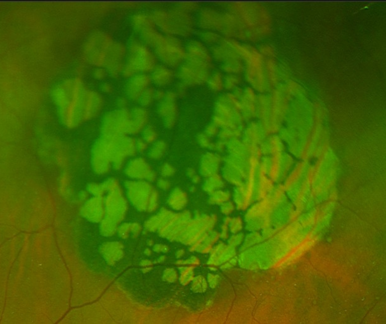
FAF shows a dense hypo-autofluorescence with lacunae showing iso-autofluorescence or hypo-autofluorescence.
Very rarely CHRPE may undergo malignant transformation, giving rise to an adenocarcinoma.
Case Examples
-
Case 1: Typical CHRPE
An asymptomatic 44 year old Asian female with best corrected visual acuity of 6/6 (20/20) in the left eye.
-
Case 2: Typical CHRPE
A 22 year old Middle Eastern female with best corrected visual acuity of 6/6 (20/20) in the right eye.
-
Case 3: CHRPE with lacunae
An asymptomatic 65 year old Caucasian female with best corrected visual acuity of 6/7.5 (20/25) in the left eye and nuclear sclerosis noted on anterior eye examination.
-
Case 4: CHRPE with lacunae
An asymptomatic 68 year old Caucasian female with best corrected visual acuity of 6/6 (20/20) in the left eye.
-
Case 5: CHRPE with subretinal cleft
An asymptomatic 35 year old Asian male with best corrected visual acuity of 6/6 (20/20) in the left eye.
-
Case 6: CHRPE with pigment clumping
An asymptomatic 60 year old Caucasian male with best corrected visual acuity of 6/6 (20/20) in the right eye.
Differential Diagnosis
References
Fung AT, Pellegrini M, Shields CL. (2014) Congenital hypertrophy of the retinal pigment epithelium: enhanced-depth imaging optical coherence tomography in 18 cases. Ophthalmology. 2014 Jan;121(1):251-256.
Shields JA, Eagle RC Jr, Shields CL, Brown GC, Lally SE. (2009) Malignant transformation of congenital hypertrophy of the retinal pigment epithelium. Ophthalmology. 2009 Nov;116(11):2213-6.