
Overview
Choroidal melanocytosis is characterised by an accumulation of heavily pigmented, but otherwise normal melanocytes within the choroid. It represents a diffuse tissue involvement with minimal impact on choroidal thickness. In contrast, a melanocytoma is a localised collection of heavily pigmented melanocytes, creating a nodular and elevated mass.
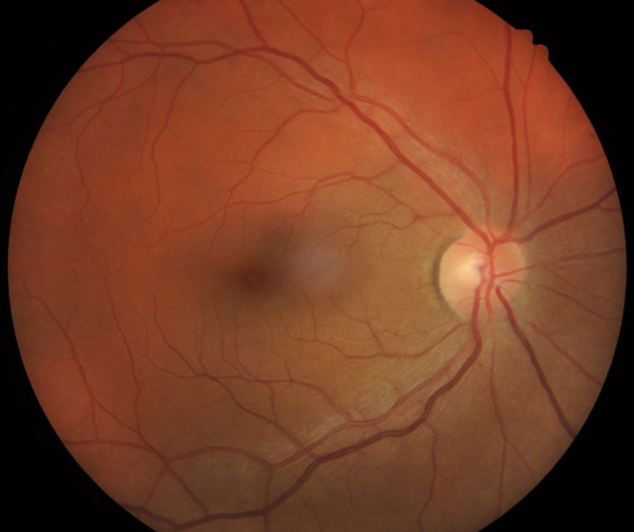
Clinically, choroidal melanocytosis presents as an area greater than 5mm in diameter of homogenous, dark brown pigmentation. OCT imaging shows the lesion to be flat.
Oculo (dermal) Melanocytosis
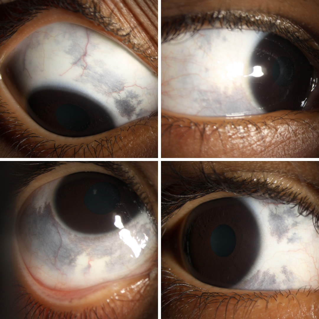
Choroidal melanocytosis falls within the spectrum of oculo (dermal) melanocytosis. Oculodermal melanocytosis typically affects the uvea and sclera as well as the choroid and may be characterised by increased pigmentation cutaneously in the periocular area (nevus of Ota – imaged here). Choroidal melanocytosis may however be isolated with no other areas of increased pigmentation.
It is estimated that one in 400 Caucasian patients with ocular melanocytosis will develop uveal melanoma. Unfortunately however, the increased fundus pigmentation in patients with choroidal melanocytosis can make it more difficult to detect a small pigmented melanoma. Augsberger et al. proposed that, based on their histological analysis of the lesions, the increased risk of melanoma in these cases is proportional to the percentage of uvea involved.
Patients with choroidal melanocytosis require annual monitoring to ensure the early detection of melanotic changes.
Case Examples
-
Case 1
An 11 year old Cacuasian male with best corrected visual acuity of 6/6 (20/20) in each eye. The left eye is unremarkable so this case will focus on the right eye.
-
Case 2
A 47 year old Caucasian female with best corrected visual acuity of 6/6 (20/20) in each eye.
Fundus photograph (1), green free (2) and red free (3) images - Right eye
More infoOptomap widefield (1), green separation (2) and red separation (3) images - Right eye
More infoFundus autofluorescence image (Right eye)
More infoSpectralis OCT line scan through the area of melanocytosis
More info
Differential Diagnosis
References
Acaba-Berrocal LA. Bekerman, VP. BS. Shields, CL (2017) Oculodermal Melanocytosis: Not to Be Overlooked: Abnormality portends guarded prognosis for choroidal melanoma. Retinal Today October 2017.
Augsburger, JJ. Trichopoulos, N. Correa, ZM. Hershberger, V. (2006) Isolated choroidal melanocytosis: A distinct clinical entity? Graefe's Arch Clin Exp Ophthalmol 244: 1522-1527.