
Overview
Ocular metastases occurs most commonly in the choroid due to the high vascular supply within this tissue. The primary site of the cancer usually arises from the breast or lung. Bilateral multifocal metastatic disease typically occurs secondary to breast cancer whereas a unilateral, solitary metastatic lesion occurs secondary to lung cancer.
Funduscopic features include a creamy white or yellow mass associated with subretinal fluid. They may appear flat, plateau or rarely, mushroom shaped. They are most commonly located posterior to the equator.
OCT imaging shows a lumpy bumpy anterior surface with an undulating and thickened RPE. This is in contrast to a smoother dome shape of a choroidal melanoma. Overlying subretinal fluid is frequently seen.
Autofluorescence imaging can show regions of hyper autofluorescence corresponding to lipofuscin or subretinal fluid.
B-scan ultrasound are acoustically dense compared to choroidal melanomas which are acoustically hollow. They also have a lower height to base ratio and can appear flat or slightly dome-shaped.
Case Examples
-
Case 1
43 year old Caucasian female with a history of breast cancer requiring multiple chemotherapy treatments.
Optomap (1), red separation (2) and green separation (3) images - right eye
More infoOptomap (1), red separation (2) and green separation (3) images - left eye
More infoSpectralis OCT line scan (right eye, inferior lesion)
More infoSpectralis OCT volume scan (right eye, inferior lesion)
More infoSpectralis OCT line scan (right eye, temporal lesion)
More infoSpectralis OCT volume scan (right eye, temporal lesion)
More infoSpectralis OCT line scan (left eye lesion)
More infoSpectralis OCT volume scan (left eye lesion)
More info -
Case 2
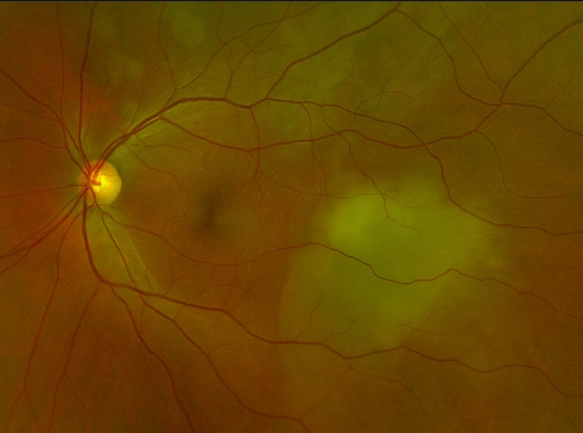
A 65 Caucasian female referred for glaucoma assessment. This case will focus on the right eye only. The patient has stage 4 lung carcinoma. Pinhole acuity 6/6+ in the right eye and Intraocular pressures 14mmHg.
Colour fundus photograph (right eye)
More infoFundus autofluoresence (FAF) imaging
More infoOptos widefield imaging (1), red separation (2) green separation (3) images
More infoCirrus OCT line scan through the nasal retina
More infoCirrus OCT line scan inferior to the optic disc
More infoCirrus OCT line scan through the area of hyper-autofluoresence
More info
Differential Diagnosis
References
Arepalli, S., Kaliki, S., & Shields, C. L. (2015). Choroidal metastases: origin, features, and therapy. Indian Journal of Ophthalmology, 63(2), 122.
Mathis, T., Jardel, P., Loria, O., Delaunay, B., Nguyen, A. M., Lanza, F., ... & Thariat, J. (2019). New concepts in the diagnosis and management of choroidal metastases. Progress in retinal and eye research, 68, 144-176.