
Overview
Choroidal osteomas are rare benign ossifying tumours, often unilateral. They may show slow growth over time and complications include serous retinal detachments, subretinal fluid, haemorrhage and choroidal neovascularisation. Tumour decalcification can also occur and is characterised by overlying RPE and choriocapillaris atrophy.
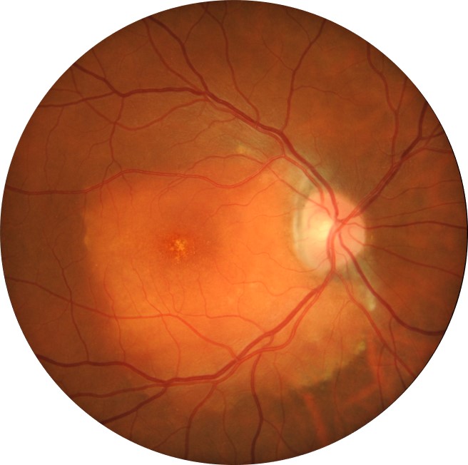
Funduscopic features include an irregularly, slightly elevated, yellow-orange choroidal mass with sharply defined or scalloped borders. They affect the juxtapapillary and macula region and can have multiple fine vascular networks on its surface.
The colour of the tumour relates to the level of RPE depigmentation. In early stages, it tends to be orange-red in colour but progress to a yellowish colour in latter stages.
OCT shows a latticework pattern with variable choroidal reflectivity. Outer retinal thinning and photoreceptor loss occurs in a decalcified choroidal osteoma.
OCT angiography can be useful in identifying a choroidal neovascular membrane.
FAF features can be variable depending on the degree of decalcification, atrophy and subretinal fluid. Generally, decalcified osteomas exhibit a reduced autofluorescence.
B-scan ultrasound shows high acoustic reflectivity (due to the calcification). A ‘pseudo optic nerve’ appearance can be seen as there is also a characteristic shadowing posterior to the tumour.
Case Examples
-
Case 1
An asymptomatic Asian male with best corrected visual acuity 6/9 (20/30) in the right eye. Grade 2 nuclear sclerosis and cortical cataracts were noted in this eye.
Fundus photograph (1), green free (2) and red free (3) images - Right eye
More infoOptomap (1), red separation (2) and green separation (3) images
More infoFundus autofluorescence image
More infoSpectralis OCT line scan through the lesion
More infoSpectralis OCT volume scan through the lesion
More infoB-scan ultrasound
More info -
Case 2
An asymptomatic 45 year old Caucasian female with best corrected visual acuity of 6/4.8 (20/15) in the right eye.
Fundus photograph (1), green free (2) and red free (3) images - right eye
More infoOptomap (1), red separation (2) and green separation (3) images
More infoFundus autofluorescence image
More infoSpectralis OCT line scan through the lesion
More infoSpectralis OCT volume scan through the lesion
More info -
Case 3
A 40 year old Caucasian male with best corrected visual acuity of 6/15 (20/50) in the left eye. He reports that this eye has always had reduced vision, compared with the right eye.
Fundus photograph (1), green free (2) and red free (3) images
More infoStereoscopic image of the lesion
More infoOptomap (1), red separation (2) and green separation (3) images
More infoFundus autofluorescence image
More infoSpectralis OCT line scan through the lesion
More infoSpectralis OCT volume scan through the lesion
More infoSpectralis OCT volume inferior to the lesion
More infoB-scan ultrasound
More info -
Case 4
A 45 year old South American female with best corrected visual acuity of 6/6 in the left eye.
Differential Diagnosis
References
Alameddine, R. M., Mansour, A. M., & Kahtani, E. (2014). Review of choroidal osteomas. Middle East African journal of ophthalmology, 21(3), 244.
Olguin-Manríquez, F., Enríquez, A.B., Crim, N. et al. (2018) Multimodal imaging in choroidal osteoma. Int J Retin Vitr 4, 30
Theodoros E. et al. (2014) Diagnosis and Monitoring of Choroidal Osteoma through Multimodal Imaging. Case Reports in Medicine Volume 2014, Article ID 393804, 4 pages